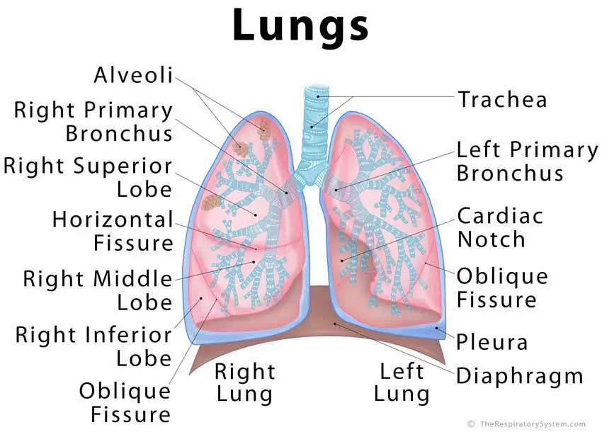
3d Illustration Of Human Body Lungs Anatomy Royalty Biology Diagrams The base of the lungs is concave to follow the contour of the diaphragm. The left lung is slightly smaller than the right lung because 2/3 of the heart is located on the left side of the body. The left lung contains the cardiac notch, an indentation in the lung that surrounds the apex of the heart. Each lung consists of several distinct lobes The current article provides a labeled diagram of the human lungs as well as a description of the parts and their functions. Lungs are an excellent example of how several tissues can be compactly arranged, yet providing a large surface area for gaseous exchange. The current article provides a labeled diagram of the human lungs as well as a Where are the Lungs Located. The lungs are located a little toward the posterior part of the human body, just below the collarbone, extending down to the diaphragm, the muscular partition that separates the chest and abdominal cavities.The left and right lungs are situated on the two sides of the body with the heart, another vital organ in the thoracic cavity, located a little in front of, and
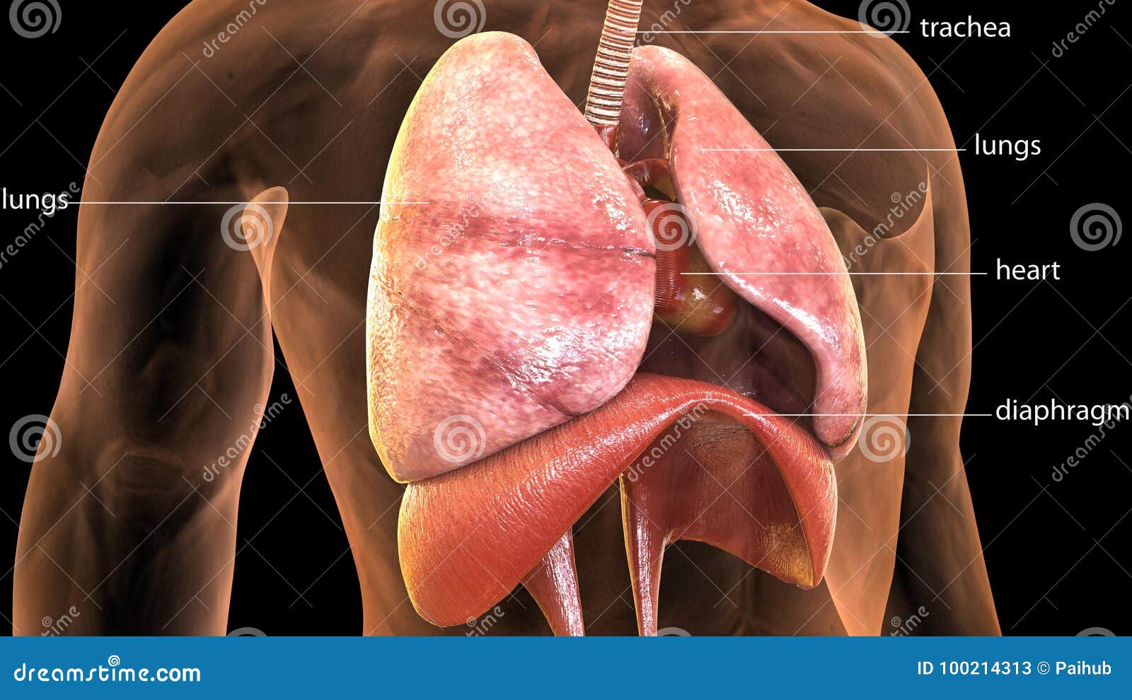
Diagram of Human Lungs shows that the lungs begin at the bottom of our trachea. The trachea is a tube that carries the air in and out of our lungs. The human lung anatomy shows paired organs that are located in the thoracic cavity of the upper body. The lungs are the primary organ of the respiratory system. It. 8 min read. Diagram of Human The anatomy of the human respiratory system begins where the air enters the body first - the nose. In addition to olfaction, the nose warms, filters and moistens in the inhaled air. Simple Lungs diagram. The lung, the human gas-exchanging structure, resides in the chest (thorax) wherein its intricate tissues are protected by the muscular

Human Lungs Anatomy Biology Diagrams
General Anatomy: diagrams showing the general organisation of the thorax with the pleural cavity and mediastinum; Anatomy of the lungs Anatomical structures of the respiratory system. 125 pulmonary anatomical structures were labeled. Those labels are grouped into subcategories, you can hide or show them on the "Anatomical parts" tab:
Anatomical Position and Relations. The lungs lie either side of the mediastinum, within the thoracic cavity. Each lung is surrounded by a pleural cavity, which is formed by the visceral and parietal pleura.. They are suspended from the mediastinum by the lung root - a collection of structures entering and leaving the lungs. The medial surfaces of both lungs lie in close proximity to several The current article provides a labeled diagram of the human lungs as well as a description of the parts and their functions. The lungs are a pair of spongy, air-filled organs located on either side of the chest (thorax). The trachea (windpipe) conducts inhaled air into the lungs through its tubular branches, called bronchi. The lungs are the main part of your respiratory system. Here is how lungs work as the center of your breathing, the path a full breath takes in your body, and a 3-D model of lung anatomy.