
A Group of Promising Proteins Biology Diagrams Keywords: cell cycle, labeled nucleosides, BrdU, EdU, markers of cell cycle phases, DNA labeling, time lapse microscopy 1. Introduction A common task of many research teams is the analysis of cell cycle progression through the distinct cell cycle phases. Various methods can be used for this purpose.
We demonstrate how to mitigate the effects of cell cycle heterogeneity in scRNA-seq data by calculating cell cycle phase scores based on canonical markers, and regressing these out of the data during pre-processing. Abstract Accurate identification of cell cycle phases in single-cell RNA-sequencing (scRNA-seq) data is crucial for biomedical research. Many methods have been developed to tackle this challenge, employing diverse approaches to predict cell cycle phases. In this review article, we delve into the standard processes in identifying cell cycle phases within scRNA-seq data and present several Do Propidium Iodide staining and so Dlow cytometry. This will tell you where your cells are being arrested in cell cycle. Now so Cyclin and CDKs of that specific phase for confirmation.
cycle markers and biosensors Biology Diagrams
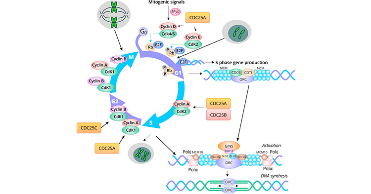
According to the complex changes in cell structure and biosynthesis, the cell cycle is divided into four phases: gap 1 (G1), DNA synthesis (S), gap 2 (G2), and mitosis (M). Determining which cell cycle phases a cell is in is critical to the research of cancer development and pharmacy for targeting cell cycle.
Here we review classical approaches that rely on cell fixation to characterise the cell-cycle status and its regulatory enzymes, and we describe the more recent development of cell-cycle markers based on genetically encoded fusions of fluorescent proteins with characteristic cell-cycle features, and of fluorescent biosensor technology to probe

What are the good markers for G2/M phase of cell cycle? Biology Diagrams
Many cellular processes are regulated by cell cycle dependent changes in protein dynamics and localization. Studying these changes in vivo requires methods to distinguish the different cell cycle stages. Here we demonstrate the use of DNA Ligase I fused to DsRed1 as an in situ marker to identify S p … These antibodies to top cell senescence markers are critical for studying cell cycle arrest in research fields like development, aging, and cancer.
