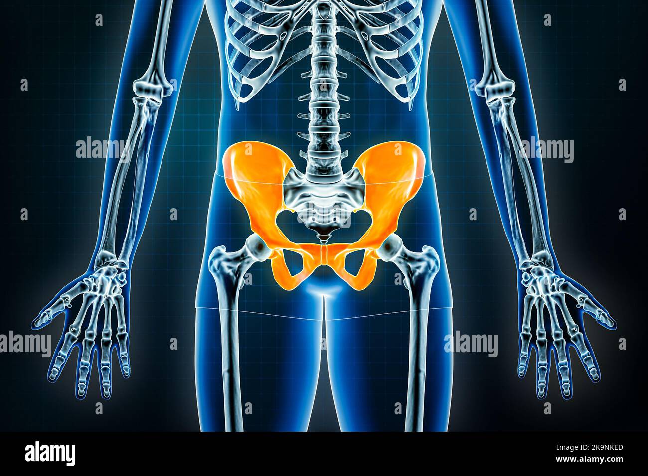
All 94 Images Pictures Of The Pelvic Bones Stunning Biology Diagrams The bony framework of the pelvis is called the pelvic girdle.It is composed of the two hip bones and the sacrum. Pelvic bones are held together by the two main joints of the pelvis; the pubic symphysis and the sacroiliac joint, and reinforced by pelvic muscles. The pelvic cavity opens superiorly to, and is continuous with, the abdominal cavity through the pelvic inlet. The bones of the pelvis are a critical part of the central portion of the skeleton. They serve as a transition from the axial skeleton and the appendicular skeleton of the lower body, serving as an attachment point for some of the strongest muscles in the human body while withstanding the forces generated by them. The curved nature of the pelvic bone creates a closed structure, itself lined

The pelvis's frame is made up of the bones of the pelvis, which connect the axial skeleton to the femurs, and therefore acts in weight bearing of the upper body. The floor of the pelvis is made up of the muscles of the pelvis, which support its contents and maintain urinary and faecal continence.

Pelvis and Perineum: Anatomy, vessels, nerves Biology Diagrams
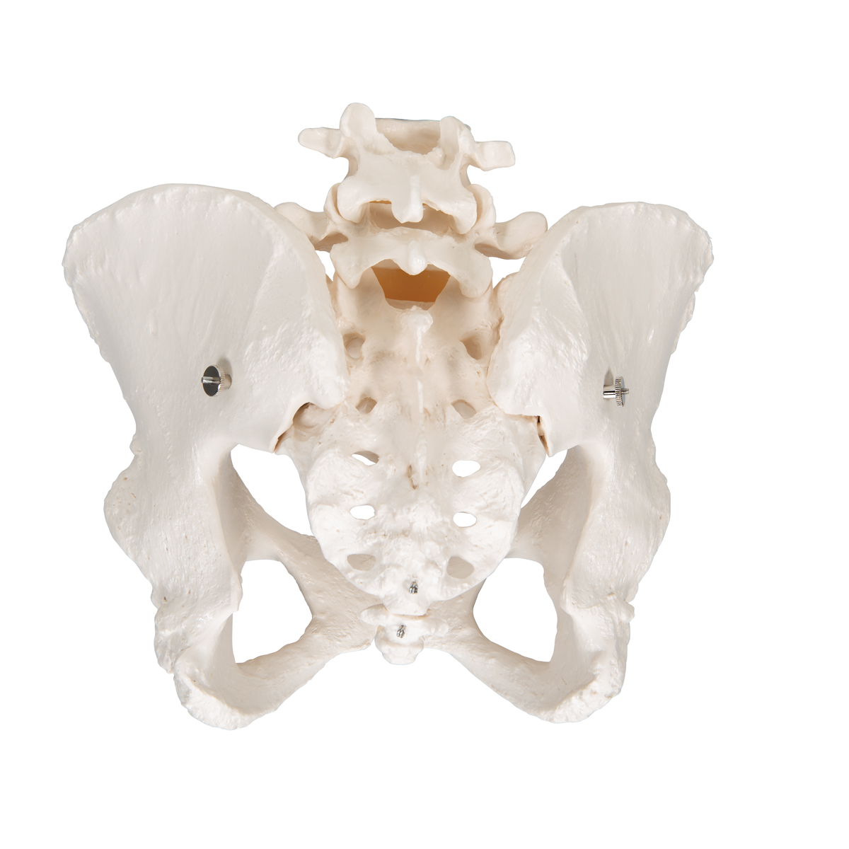
Anatomy The bony pelvis. The bony pelvis is made up of two pelvic bones - the sacrum and the coccyx. Each pelvic bone (hip bone) is made by the combination three bones namely, the ilium, pubis, and ischium. Principles of Human Anatomy (15th ed.). Wiley. ISBN 978-1119662686. Standring, S. (2018). Gray's Surgical Anatomy (1st ed
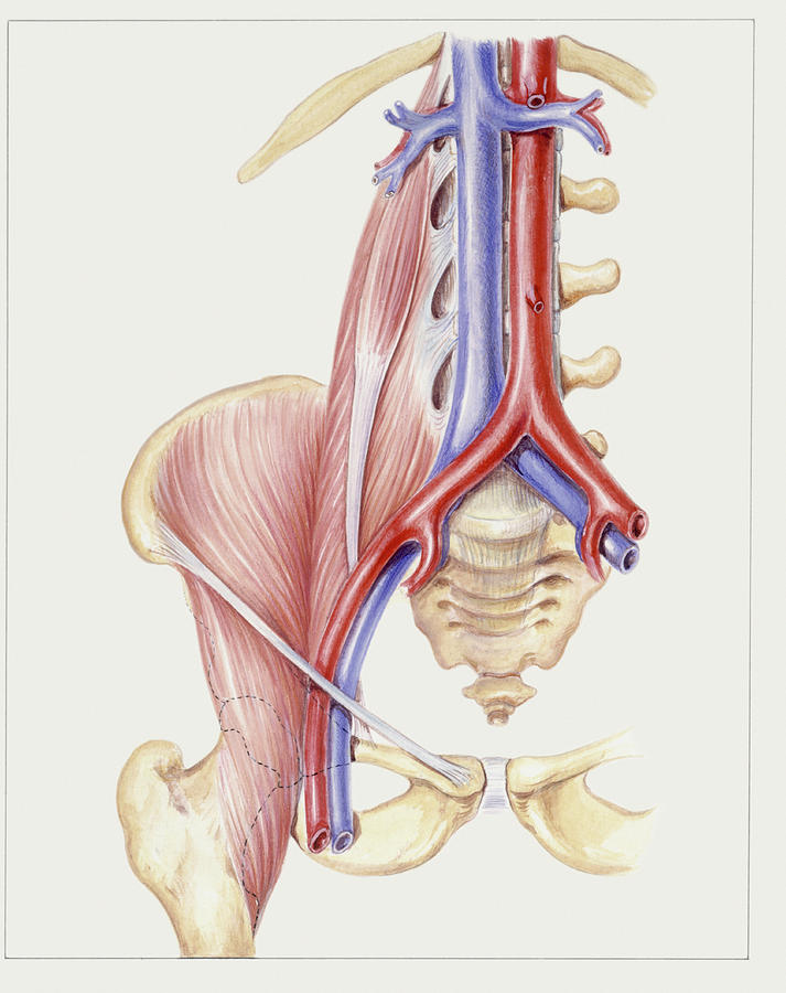
The pelvic floor is a unique anatomical location where the balance of the different pressures, either visceral, muscular, or liquid play a fundamental role in the physiological functioning of all the structures contained therein. The pelvis is bounded superiorly by the imaginary line between the pubis and sacral promontory and inferiorly as the line between the ischial tuberosity and the apex Responsible for supporting upper body weight, the pelvis is defined as the middle part of the human body between the lumbar region of the abdomen superiorly and thighs inferiorly. The human pelvis is composed of the bony pelvis, the pelvic cavity, the pelvic floor, and the perineum. In addition to carrying upper body weight, this multi-surfaced girdle can transfer upper body weight to the
Wikipedia Biology Diagrams
Your pelvis is the bony structure inside your hips, buttocks and pubic region. It's the seat that holds up your upper body when you sit, stand or walk. The hole in the middle of your pelvis serves as the birth canal during vaginal delivery. Your pelvic anatomy can shift to accommodate childbirth. The same human pelvis, front imaged by X-ray (top), magnetic resonance imaging (middle), and 3-dimensional computed tomography (bottom). The pelvis (pl.: pelves or pelvises) is the lower part of an anatomical trunk, [1] between the abdomen and the thighs (sometimes also called pelvic region), together with its embedded skeleton [2] (sometimes also called bony pelvis or pelvic skeleton).