
Anatomy and Physiology of Salivary Glands Biology Diagrams salivary gland, any of the organs that secrete saliva, a substance that moistens and softens food, into the oral cavity of vertebrates.. Salivary glands may be predominantly serous, mucous, or mixed in secretion. Mucus is a thick, clear, and somewhat slimy substance. Serous secretion is a more liquid opalescent fluid composed of water and proteins, such as the digestive enzyme amylase. The salivary glands are exocrine glands which produce a digestive fluid called saliva.They are accessory organs of the digestive system and are positioned in the head, in and around the oral cavity and secrete their salivary contents into the mouth.. The salivary glands are divided into the major and minor salivary glands: The major glands include the parotid, submandibular and sublingual glands.

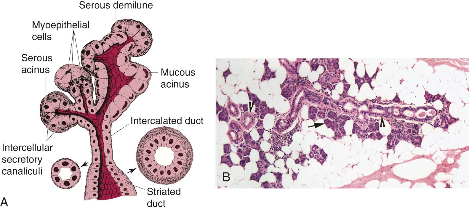
Anatomy. The glands responsible for the production of saliva include the parotid gland, the largest of the salivary glands, the submandibular glands, and the sublingual glands. The structure of the salivary glands consists of a series of ducts that eventually end in either a spherical or tubular secretory acini or end piece.

Salivary Glands: Function, Location & Anatomy Biology Diagrams
Salivary glands are classified as exocrine, and as such, they produce secretions (i.e., saliva) onto an epithelial surface via a system of ducts. Saliva secretion and production are mediated by the autonomic nervous system (ANS) and thus; salivary glands have both parasympathetic and sympathetic innervation. The salivary glands are exocrine glands that make, modify and secrete saliva into the oral cavity. They are divided into two main types: the major salivary glands, which include the parotid, submandibular and sublingual glands, and the minor salivary glands, which line the mucosa of the upper aerodigestive tract and the overwhelming entirety of the mouth [1].
Keywords. Saliva; Salivary glands; Saliva secretion; FormalPara Objectives . This contribution aims to show the anatomical and physiological characteristics of the salivary glands as entity for the production of saliva, and to present the composition of the saliva fluid as a protective medium for the mouth, the start of digestion and as diagnostic medium. Salivary gland regeneration has the potential to permanently restore salivary gland secretory function in patients with hyposalivation to improve their oral health and quality of life. The three main approaches that have been proposed are 1) gene therapy, by using viral vectors, 2) stem/progenitor cell-based therapy, and 3) replacement with a

Anatomy, Head and Neck, Salivary Glands Biology Diagrams
Your salivary glands lubricate your mouth, help you swallow, aid in digestion and help protect your teeth against harmful bacteria. You have three major types of salivary glands, including your sublingual, submandibular and parotid. Common symptoms of salivary gland disorders include fever, headaches and a lump in your cheek or under your chin. Salivary glands are exocrine glands responsible for producing and secreting saliva into the oral cavity.[8] Saliva aids in moistening food, digestion, lubrication, and oral hygiene. These glands are composed of serous, mucous, or mixed secretory cells that release enzymes, electrolytes, and mucus to maintain oral health and digestive function. Location Salivary glands are located in
