
ANATOMY OF VENTRICLES OF BRAIN Biology Diagrams jack0m / DigitalVision Vectors / Getty Images. The ventricles of the heart function to pump blood to the entire body. During the diastole phase of the cardiac cycle, the atria and ventricles are relaxed and the heart fills with blood. During the systole phase, the ventricles contract pumping blood to the major arteries (pulmonary and aorta).The heart valves open and close to direct the flow of
This detailed anatomical illustration presents a cross-sectional view of the human heart, highlighting its major chambers, valves, and blood vessels through a modern, clear design. The diagram effectively uses color coding to distinguish between oxygenated (red) and deoxygenated (blue) blood flow paths, making it an excellent educational resource for understanding cardiac anatomy. The definition of heart ventricles can be summed up as the large, lower chambers of the fibromuscular organ that work to keep blood moving through the body. Although all parts of the heart work together to carry out its daily function, the ventricles have an enormous role in maintaining adequate cardiac output to keep blood flowing. The heart works continuously from the 4th gestational week

Heart Anatomy: Complete Guide with Parts, Names & Diagram Biology Diagrams
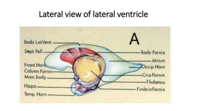
Overview Function Anatomy Conditions and Disorders Care Additional Common Questions. Overview Your heart is a muscular organ that pumps blood to your body. plural atria) and two on the bottom (ventricles), one on each side of your heart. Right atrium: Two large veins deliver oxygen-poor blood to your right atrium. The superior vena cava Anatomy . The pair of lateral ventricles are the largest of the four ventricles in the brain. They are located in the largest part of the brain, the cerebrum. The third ventricle is in the diencephalon, located in the center of the brain. The fourth ventricle is located in the hindbrain.

Anatomy. The heart is a four-chambered muscular organ with a complex structure that allows it to efficiently pump blood throughout the body. Structure & Function of the Body (17th ed.). Elsevier. ISBN 9780323597791. It is approximately the size of a fist and consists of four chambers: two atria and two ventricles. The heart is composed This human heart model comprehensively explores its intricate anatomy, including ventricles with their valves, atria with their valves, and the significance of these valves. It also examines the myocardial composition, endocardium, and the protective pericardium. The model details the main artery (aorta) and its relationship to the pulmonary artery and veins, as well as the vena cava and its A ventricle is one of two large chambers located toward the bottom of the heart that collect and expel blood towards the peripheral beds within the body and lungs. The blood pumped by a ventricle is supplied by an atrium, an adjacent chamber in the upper heart that is smaller than a ventricle.Interventricular means between the ventricles (for example the interventricular septum), while
