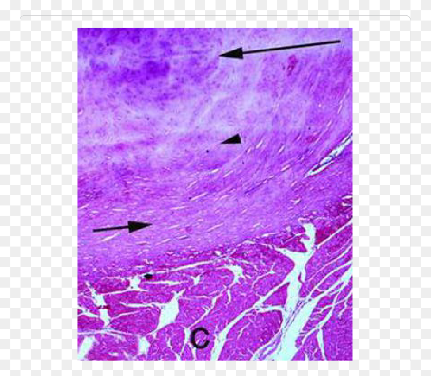
Applied Compressive Strain Governs Hyalinelike Biology Diagrams A: Perichondrium lines the outside of the hyaline cartilage and acts like a capsule it is DCCTRA. B: Chondroblast layer are transformed fibroblasts that are starting to enlarge and synthesize their own cartilage matrix. These cells are much smaller and thinner than the fully mature chondrocytes. C: Chondrocytes completely surrounded by matrix. Hyaline cartilage, elastic cartilage, and fibrocartilage are three types of cartilage with distinct properties and functions. Hyaline cartilage is the most prevalent type, characterized by its translucent appearance and smooth surface. In contrast, elastic cartilage contains a network of elastic fibers that provide flexibility. The fibrocartilage provides flexibility, toughness, and elasticity to the structural parts of the body. It contains intermediate characteristics between the dense connective tissue and the hyaline cartilage. The types of cells that occur in the fibrocartilage are fibroblasts, fibrocytes, chondroblasts, and chondrocytes.
Cartilage is a strong, flexible connective tissue that protects your joints and bones. It absorbs impacts and reduces friction between bones throughout your body.

Hyaline Cartilage: Definition, Function, Quiz Biology Diagrams
Hyaline cartilage is present in the growth plates of developing bones, aiding in their growth and development. Although hyaline cartilage has some degree of flexibility, it is relatively weaker compared to other types of cartilage, such as elastic cartilage or fibrocartilage. Fibrocartilage is seen in the glenoid labrum, acetabular labrum, menisci, pubic symphysis, etc. Also, in the places where the ligaments and tendons attach to bone. Hyaline Cartilage It is a translucent cartilage that is present on many bone joints. Hyaline cartilage is a firm structure that has collagen.

Hyaline cartilage is a type of connective tissue found in areas such as the nose, ears, and trachea of the human body. The word hyaline means "glass-like", and hyaline cartilage is a glossy, greyish-white tissue with a uniform appearance. It is one of the three types of cartilage; the other two types are elastic cartilage and fibrocartilage. Cartilage In additional to bone, cartilage is also an important part of the skeletal system creating structure, connecting bones, cushioning joints (articulations), protecting the ends of bones, and providing shock absorption. There are three different types of cartilage in the human body: hyaline cartilage, elastic cartilage, and fibrocartilage (see table below). For example, hyaline

TeachMePhysiology Biology Diagrams
Examine this slide of fibrocartilage, noting the distinctive features of fibrocartilage when compared to hyaline and elastic cartilages (see table on previous page 2_2 and the images below). Elastic cartilage resembles hyaline cartilage somewhat in histological appearance with a few subtle differences. Cartilage is a flexible connective tissue that is stiffer than most other connective tissue types but flexible enough to withstand compressive forces.