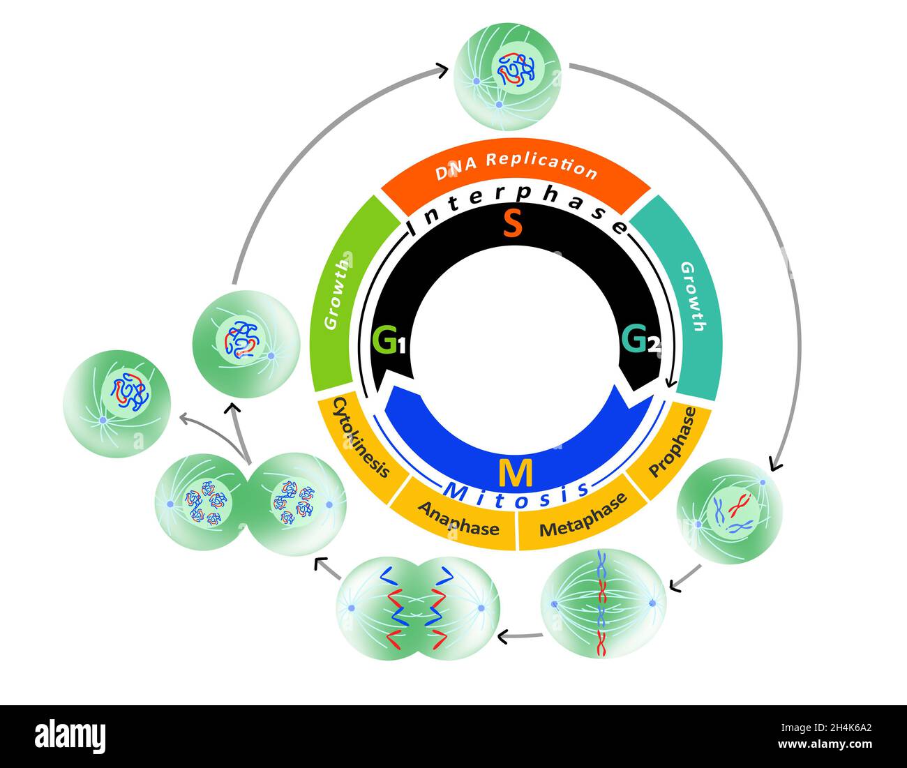
Cell cycle diagram Phases of the cell cycle and mitosis DNA Biology Diagrams The eukaryotic cell cycle consists of four distinct phases: G 1 phase, S phase (synthesis), G 2 phase (collectively known as interphase) and M phase (mitosis and cytokinesis). M phase is itself composed of two tightly coupled processes: mitosis, in which the cell's nucleus divides, and cytokinesis, in which the cell's cytoplasm and cell membrane divides forming two daughter cells. The M-Phase, also known as the Mitotic Phase, is a stage in the cell cycle where cell division occurs. It is divided into several stages: Prophase: In this stage, the chromatin condenses into a highly ordered structure called chromosomes. The nuclear envelope breaks down and spindles start to form. Prometaphase: The nuclear envelope is completely broken down, and the chromosomes are free in
The cell cycle consists of distinct phases that guide cellular activities from one stage to the next. Each phase has specific functions and checkpoints to maintain genomic integrity and prevent errors. G1 Phase (Cell Growth) The G1 phase, often referred to as the first gap phase, is a period of cellular growth and metabolic activity. We learned that a critical part of the cell cycle is the M phase, in which mitosis and cytokinesis occur. Mitosis is the process of duplicated chromosomes being aligned, separated, and moving into two new, identical daughter nuclei. Today we will dive into the details of mitosis and cytokinesis. Before we can learn about how chromosomes move
Molecular Biology of the Cell Biology Diagrams
It is the first phase of the cell cycle, recognized by the growth period where the chromosome gets duplicated as the cell prepares for division. Interphase happens between one cell division or mitotic (M) phase and the next. It is the longest part of the cell cycle involving three sub-phases. The typical duration of this phase is 23 hours.
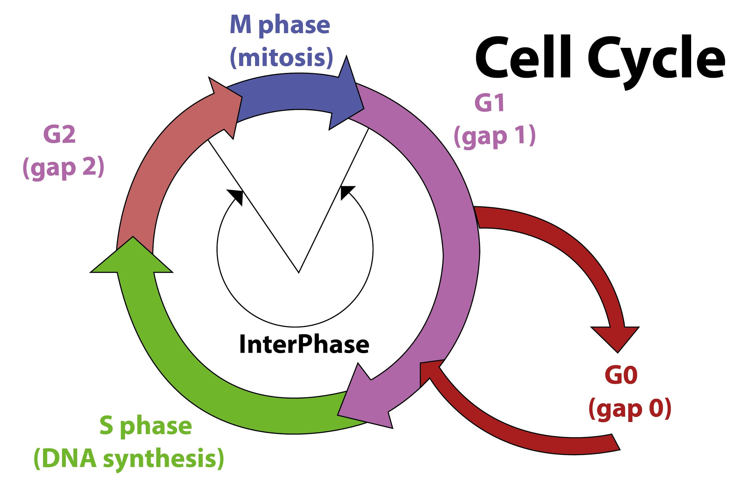
The cell cycle consists of a timed sequence of events that occur during interphase and mitosis (M). Interphase is made up of the G 1 (G = gap) phase, the S (synthesis) phase, and the G 2 phase (Fig. 15-2).Both G phases contain checkpoints that govern whether the cell moves into DNA replication (G 1 checkpoint) or into mitosis (G 2 checkpoint).. The G 1 and G 2 phases involve the synthesis of

Cell Cycle M Phase - an overview Biology Diagrams
In most animal cells, M phase takes only about an hour—a small fraction of the total cell-cycle time, which often lasts 12-24 hours. The rest of the cycle is occupied by interphase . Under the microscope, interphase appears as a deceptively uneventful interlude, in which the cell simply continues to grow in size. Overview of the Cell Cycle Phases. The two broad phases of the cell cycle are interphase and mitosis. During interphase, cells grow, replicate their DNA and organelles, and prepare for division. Interphase steps are the first gap phase (G 1), the synthesis phase (S), and the second gap phase (G 2). Cells divide during mitosis (M). The M phase of a cell cycle is also called mitosis. This is a form of asexual cell reproduction in eukaryotes, equivalent in most respects to binary fission in prokaryotes. In includes prophase, prometaphase, metaphase, anaphase and telophase, and it relies on the mitotic spindle at each cell pole.