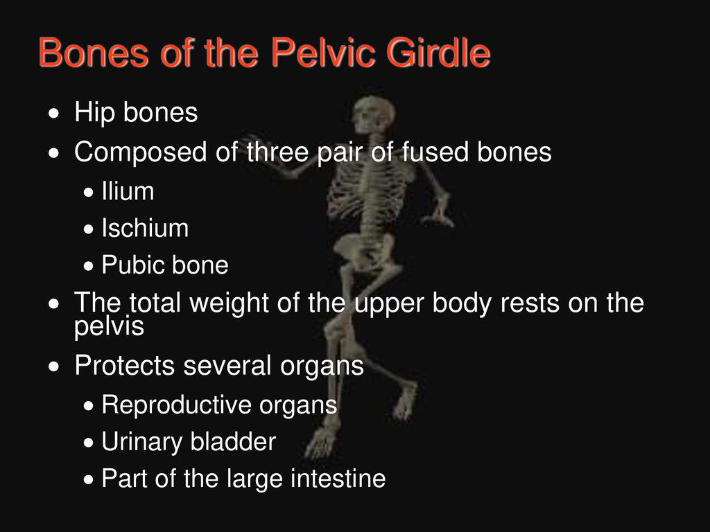
Chapter 5 The Skeletal System Biology Diagrams The pelvic bones are: . Sacrum; Coccyx; Two innominate bones, which consist of the: Ischium; Ilium; Pubis; The inlet to the pelvic canal is at the level of the sacral promontory and superior aspect of the pubic bones.; The outlet is formed by the pubic arch, ischial spines, sacrotuberous ligaments, and the coccyx. The enclosed space between the inlet and outlet is called the true pelvis, with The pelvic girdle performs several important functions in the human body. It supports the weight of the upper body, stabilizes it and transmits this weight to the lower limbs, allowing a range of actions to occur (e.g. sitting, standing, bipedal gait).It also protects the abdominopelvic viscera and provides attachment points for the smaller muscles and ligaments of the pelvic floor and the
Pelvis, in human anatomy, basin-shaped complex of bones that connects the trunk and the legs, supports and balances the trunk, and contains and supports the intestines, the urinary bladder, and the internal sex organs. Learn more about the pelvis in this article. Also known as: bony pelvis, hip girdle, pelvic bone. Written and fact-checked Structure of the Pelvic Girdle. The bony pelvis consists of the two hip bones (also known as innominate or pelvic bones), the sacrum and the coccyx.. There are four articulations within the pelvis: Sacroiliac joints (x2) - between the ilium of the hip bones, and the sacrum Sacrococcygeal symphysis - between the sacrum and the coccyx. Pubic symphysis - between the pubis bodies of the two Pelvic Girdle In human anatomy, Pelvic Girdle is a basin-shaped structure of bones that connects the trunk of the legs and holds the support of the intestines, urinary bladder, and the interior sex organs. It is also termed as the bony pelvis. Pelvic Girdle Bones and Structure The Pelvic Girdle consists of two paired hipbones,

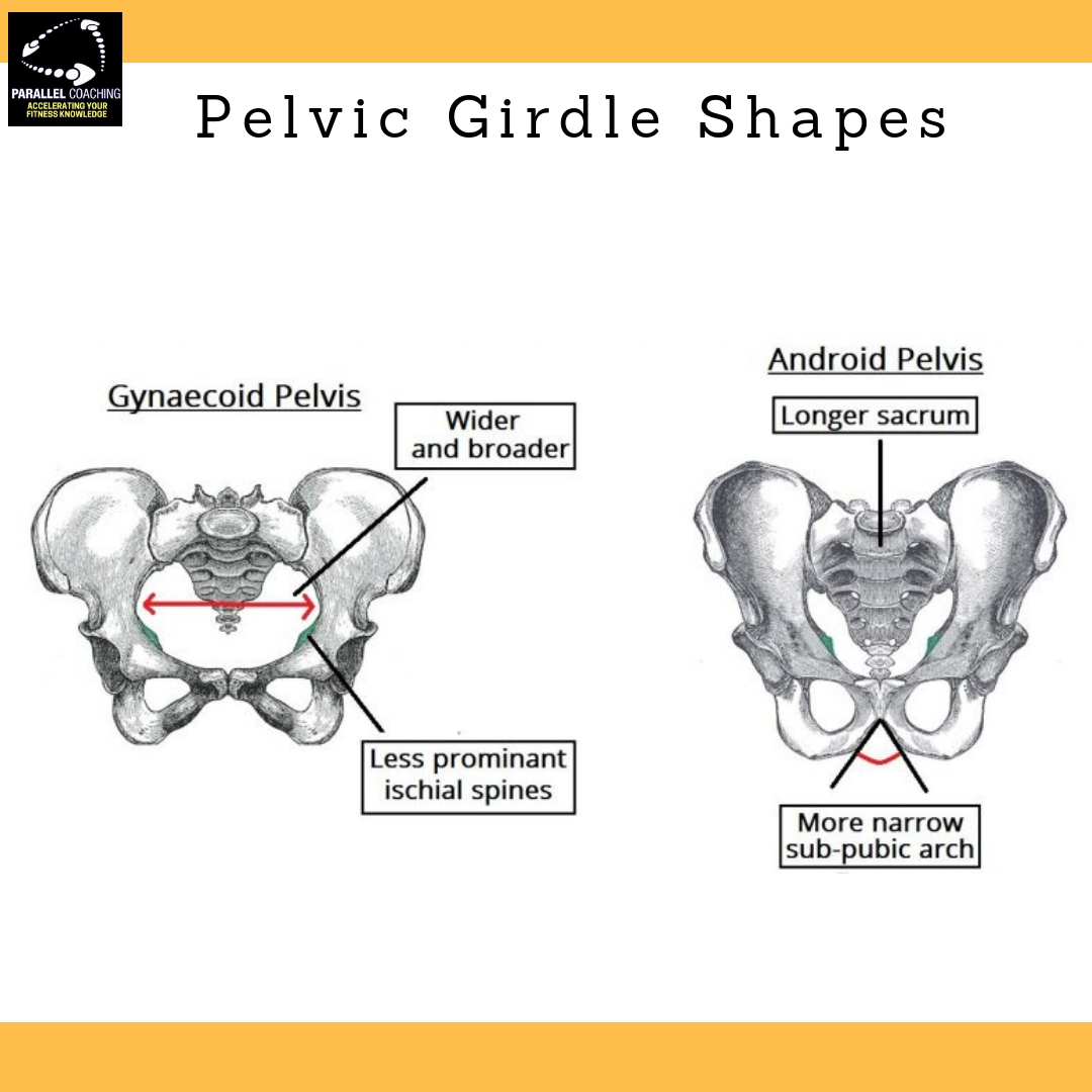
Pelvic Girdle Anatomy Biology Diagrams
The pelvic girdle (hip girdle) is formed by a single bone, the hip bone or coxal bone (coxal = "hip"), which serves as the attachment point for each lower limb. Each hip bone, in turn, is firmly joined to the axial skeleton via its attachment to the sacrum of the vertebral column. The right and left hip bones also converge anteriorly to attach to each other. The pelvic girdle (hip girdle) is formed by a single bone, the hip bone or coxal bone (coxal = "hip"), which serves as the attachment point for each lower limb.Each hip bone, in turn, is firmly joined to the axial skeleton via its attachment to the sacrum of the vertebral column. The right and left hip bones also converge anteriorly to attach to each other. The two hip bones (also called coxal bones or os coxae) are together called the pelvic girdle (hip girdle) and serve as the attachment point for each lower limb. When the two hip bones are combined with the sacrum and coccyx of the axial skeleton, they are referred to as the pelvis.The right and left hip bones also converge anteriorly to attach to each other at the pubic symphysis (Figure 8.3.1).