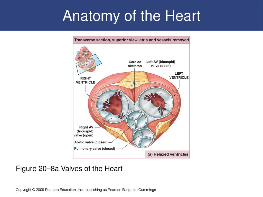
Concise Medical Knowledge Biology Diagrams These are high-frequency sounds and arise from mitral and tricuspid valve Tricuspid valve The valve consisting of three cusps situated between the right atrium and right ventricle of the heart. Heart: Anatomy closure (S1), as well as aortic and pulmonary valve Pulmonary valve A valve situated at the entrance to the pulmonary trunk from the Understanding heart valves anatomy is important in grasping the overall function of the heart.The heart is one of the most important organs in the body.It is responsible for propelling blood to every organ system, including itself.Other articles have discussed at length the gross anatomy of the heart and its four chambers. Special mention has also been made of the fact that the heart has a

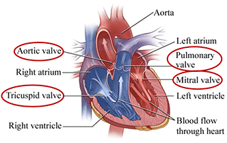
Anatomy of the heart and blood flow (video) Basics about S3, S4, and heart murmurs; Don't forget to take the heart sounds quiz when you are done reviewing Memorize the information below because it is crucial in understanding heart sounds and which valve represents each sound. The atrioventricular valves are located between the atria and the ventricles. They close during the start of ventricular contraction (systole), producing the first heart sound. There are two AV valves: Tricuspid valve - located between the right atrium and the right ventricle (right atrioventricular orifice). It consists of three cusps What are the symptoms of heart valve problems? Some people can have a heart valve condition but not have any symptoms at all. Heart valve conditions tend to get worse over time, so symptoms may appear as a person gets older. The sound of your heartbeat is the sound of your heart valves opening and closing. The first sign of a heart valve

What Are Heart Sounds and How to Know If They're Normal Biology Diagrams
Basics of Heart Sounds - S1 and S2 heart sounds. There are 2 main heart sounds that can be heard during auscultation: S 1 and S 2, also affectionately known as 'lub' and 'dub' respectively.. The S 1 and S 2 heart sounds are part of the normal heart sounds. Source: University of Michigan Murmur library S 1 heart sound corresponds to the closing of the mitral and tricuspid valves
These sounds, primarily the "lub-dub" generated by the closure of heart valves, serve as a diagnostic tool for assessing heart health. The "lub," or first heart sound (S1), corresponds to the closure of the atrioventricular valves, signaling the onset of ventricular systole. Vibrations from closing the two valves known as the mitral and tricuspid valves cause the first "lub" heart sound. These valves close to prevent blood from flowing into either atrium after the

Concise Medical Knowledge - Lecturio Biology Diagrams
Heart sounds occur from the closing of heart valves, just like a door slamming shut, and the sound is transmitted in the direction of blood flow. There are four valves in the heart: two atrioventricular (AV) valves and two semilunar valves. The two atrioventricular valves separate the atria from the ventricles and include the tricuspid valve on