Coronary Arteries And Heart Anatomy Worksheet Biology Diagrams The coronary artery branches are the first of many branches off your aorta. Coronary artery structure. There are two coronary arteries, each containing several branches: Right coronary artery (RCA): The RCA supplies blood to your right atrium and right ventricle (where deoxygenated blood goes before heading to the lungs). Its branches supply Coronary arteries. The coronary arteries arise from the root of the ascending aorta.Recall that the aortic valve has three semilunar cusps, also known as the sinuses of Valsalva. The left and right semilunar cusps give rise to the corresponding left and right coronary arteries (respectively). The third sinus - which is the posterior semilunar cusp - is not associated with a coronary vessel Smaller branches of the coronary arteries include: obtuse marginal (OM), septal perforator (SP), and diagonals. Why are the coronary arteries important? Since coronary arteries deliver blood to the heart muscle, any coronary artery disorder or disease can have serious implications by reducing the flow of oxygen and nutrients to the heart muscle.

Anatomy. The coronary arteries arise from the sinuses of Valsalva, just past the origin of the aortic root. The right coronary artery (RCA), arising from the anterior aortic sinus, supplies blood to the right atrium, right ventricle, sinoatrial node, atrioventricular (AV) node, and select portions of the left ventricle. The coronary arteries are the arterial blood vessels of coronary circulation, which transport oxygenated blood to the heart muscle.The heart requires a continuous supply of oxygen to function and survive, much like any other tissue or organ of the body. [1]The coronary arteries wrap around the entire heart. The two main branches are the left coronary artery and right coronary artery. Coronary artery disease. The coronary arteries are essentially functional end arteries, though some anastomoses exist at the arteriolar level. Anastamoses can potentially occur between the arteries within the atrioventricular groove, the interventricular and conus branches. In slow occlusion, arterioles may develop between the branches described.

Coronary anatomy and anomalies Biology Diagrams
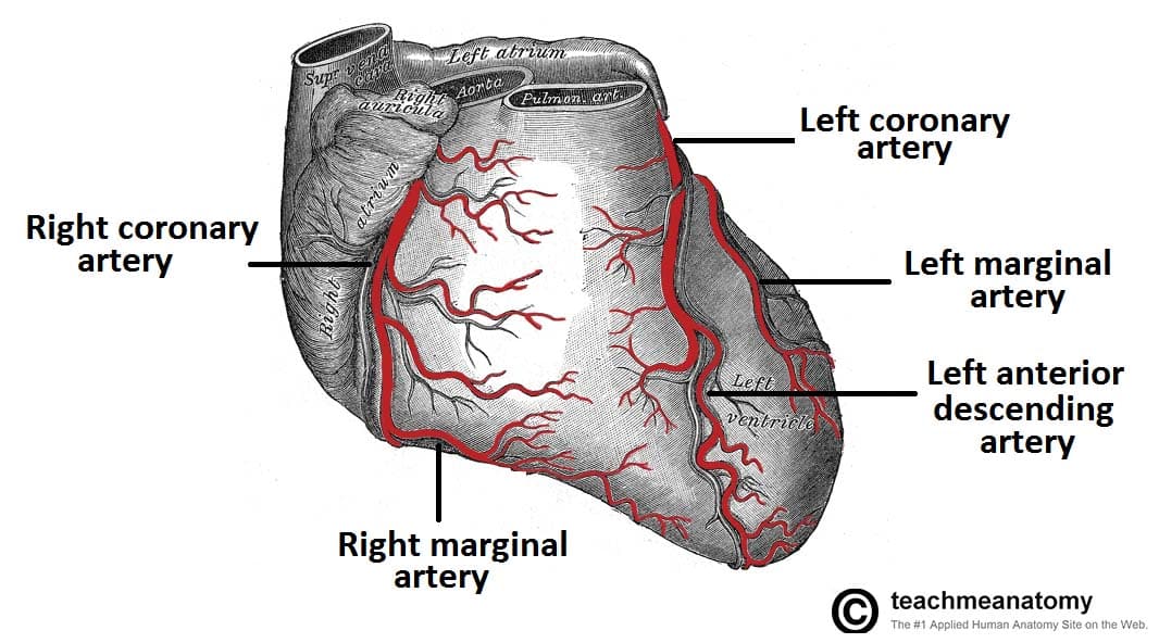
In this article we describe the anatomy of the coronary arteries of the heart and some of the anomalies with illustrations and CT-images. The right coronary artery arises from the anterior sinus of Valsalva and courses through the right atrioventricular (AV) groove between the right artium and right ventricle to the inferior part of the Naming Coronary Arteries. There are two main coronary arteries which branch to supply the entire heart. They are named the left and right coronary arteries, and arise from the left and right aortic sinuses within the aorta. The aortic sinuses are small openings found within the aorta behind the left and right flaps of the aortic valve.When the heart is relaxed, the back-flow of blood fills Gross anatomy. The typical configuration consists of two coronary arteries, a left main coronary artery (LMCA) and a right coronary artery (RCA), arising from the left (posterior) and right (anterior) aortic or coronary sinuses, respectively, in the proximal ascending aorta.These are the only two branches of the ascending aorta. The right coronary artery courses in the right atrioventricular