Craniosynostosis Biology Diagrams This study aimed to establish a comprehensive quantitative framework for accurately capturing the cranial suture and fontanelle morphologies in infants. A total of 69 CT scans of 2-4 month‐old infant heads were segmented to identify semilandmarks at the borders of cranial sutures and fontanelles.
A total of 69 CT scans of 2-4 month‐old infant heads were segmented to identify semilandmarks at the borders of cranial sutures and fontanelles. Morphological characteristics, including length, width, sinuosity index (SI), and surface area, were measured. For this, an automatic method was developed to determine the junction points between
Sutures and Fontanelles Biology Diagrams
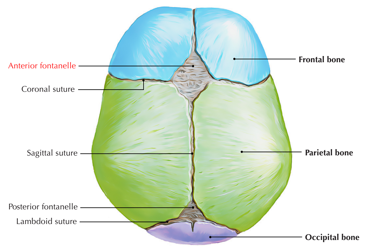
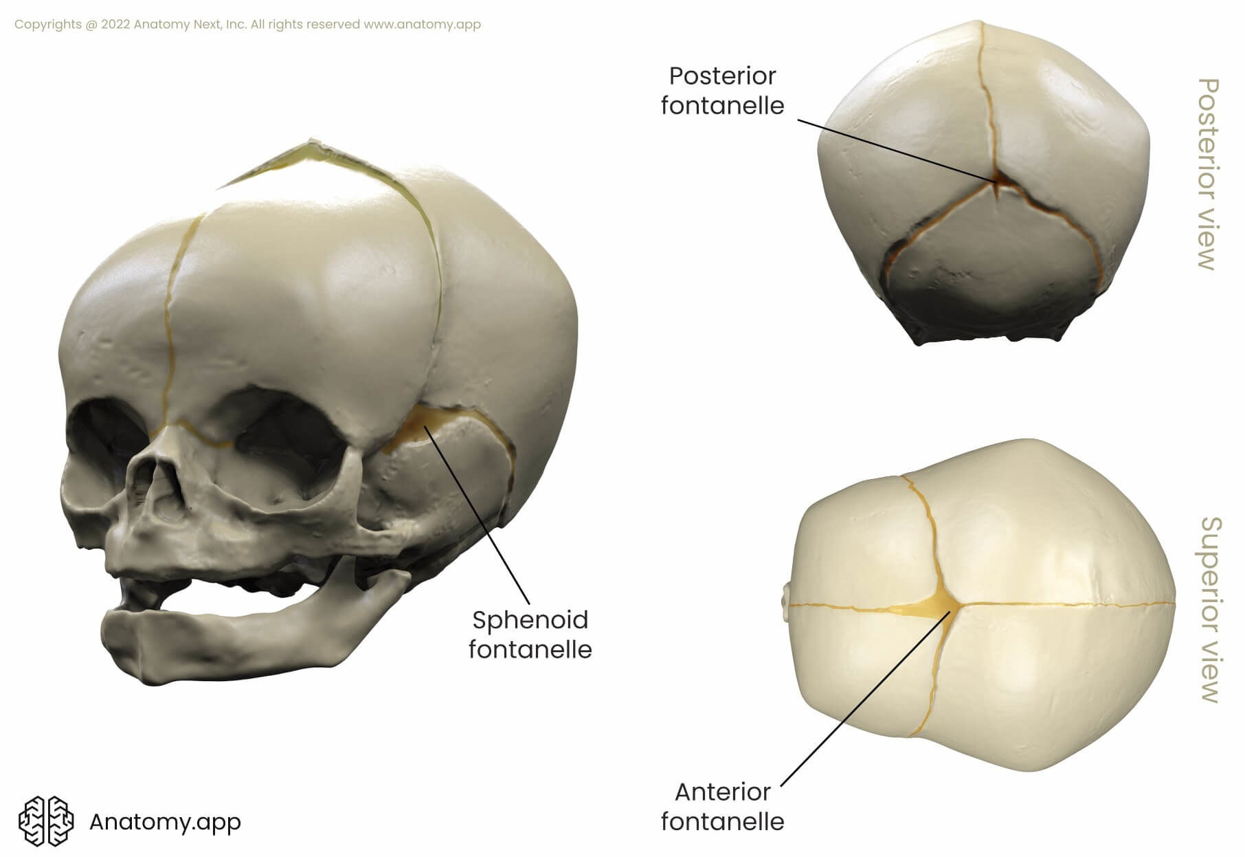
The sutures of the skull, also referred to as the cranial sutures, are fibrous joints that connect the bones of the skull.They appear as intricate thin lines that mark the adherence between the bones and the growth and closure of the cranial fontanelles. The dense fibrous tissue that connects the sutures is made mostly out of collagen. These joints are fixed, immovable, and they have no cavity. Without flexible sutures and fontanelles, the child's brain could not grow enough. The child would develop brain damage. Feeling the cranial sutures and fontanelles is one way that health care providers follow the child's growth and development. They are able to assess the pressure inside the brain by feeling the tension of the fontanelles.

Cranial sutures and fontanels. Print. Sections. Products and services. Joints made of strong, fibrous tissue (cranial sutures) hold the bones of your baby's skull together. The sutures meet at the fontanels, the soft spots on your baby's head. The sutures remain flexible during infancy, allowing the skull to expand as the brain grows. At birth, the newborn's skull consists of five major bones (two frontal, two parietal, and one occipital) that are separated by connective tissue junctions known as cranial sutures.[1] The sutures function as seams, and they are highly necessary to facilitate the movement and molding of the cranium through the birth canal during labor. They also allow for rapid postnatal growth and development 10.1055/b-0034-87890 Sutures and Fontanelles The width of the sutures is highly variable in neonates; therefore, measurements are not reliable. Plain film, ultrasound (US), and computed tomography (CT) can be used for assessment of sutural patency. The coronal suture is the first to manifest widening in response to increased intracranial pressure (ICP; upper limit of normal…

Anatomy, Head and Neck: Fontanelles Biology Diagrams
Why are Fontanelles and cranial sutures important? Both are very important as they promote brain growth. How? Because brain expands faster than the surrounding bones do, having flexible sutures and membraneous areas (fontanelles) in diverse places of the cranial vault will allow the rapid stretching of the cranium, while brain volume increases.

The human cranium presents an intricate network of sutures and fontanelles that are crucial for both development and structural integrity. From the superior view, these anatomical landmarks provide essential insights into cranial growth patterns, potential pathologies, and developmental milestones. Understanding these features is fundamental for medical professionals, particularly in