
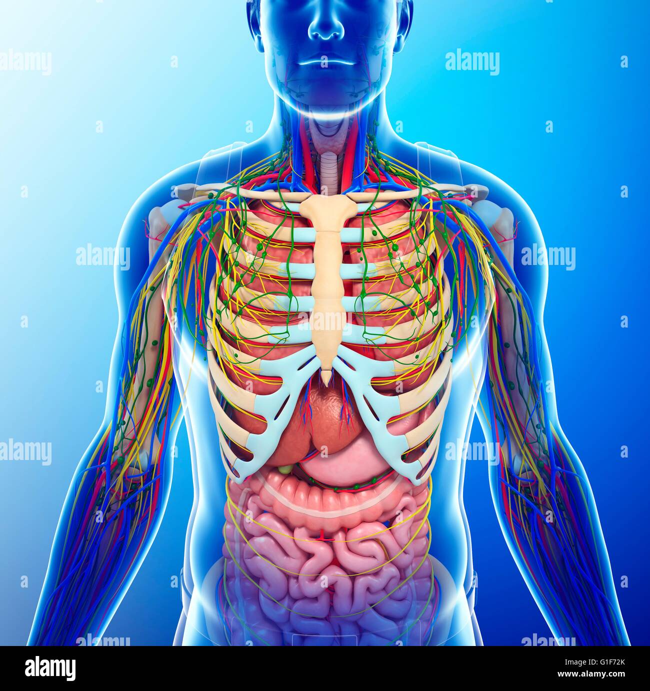
DIAGRAM Female Chest Diagram Biology Diagrams thoracic cavity, the second largest hollow space of the body. It is enclosed by the ribs, the vertebral column, and the sternum, or breastbone, and is separated from the abdominal cavity (the body's largest hollow space) by a muscular and membranous partition, the diaphragm.It contains the lungs, the middle and lower airways—the tracheobronchial tree—the heart, the vessels transporting The thorax, commonly known as the chest, is a vital region of the human body that provides protection to critical organs, including the heart and lungs, Sobotta Atlas of Human Anatomy: Volume 2 - Thorax, Abdomen, and Pelvis (14th ed.). Urban & Fischer. ISBN 978-0723434521.
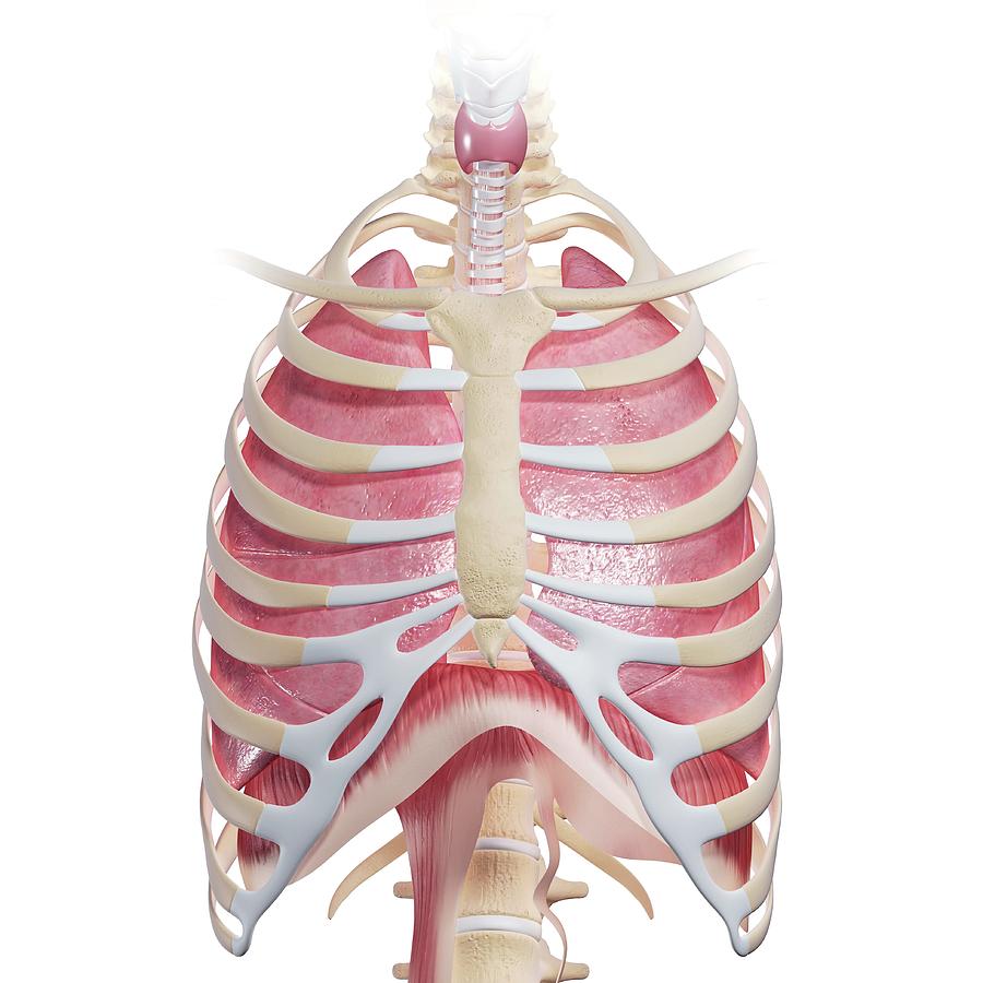
The thorax is the area of the body situated between the neck and the abdomen. The thorax itself can be split up into various areas that contain important structures.. The thorax is bound by bony structures including the 12 pairs of ribs and thoracic vertebrae, whilst also being supported by many ligaments and muscles.. The muscles of the thorax are also important for the vital actions of Atlas of anatomy of the human body: anatomical illustrations of the lungs, chest, bronchi, trachea and thoracic lymph nodes Human anatomy atlas IMAIOS DICOM Viewer Free DICOM viewer vet-Anatomy Anatomy of the chest and the lungs: anatomical illustrations. IMAIOS CGA 2023.

Description, Anatomy, & Physiology Biology Diagrams
Diaphragm Anatomy. The diaphragm is a dome-shaped muscle that forms the floor of the thoracic cavity, playing a fundamental role in the mechanics of breathing. This intricate network highlights the chest cavity's role in maintaining the body's oxygen supply and overall circulatory health. Nerve Innervation.
Your thoracic cavity is the large space in your chest where some of your body's most important work gets done. If you're interested in learning about the thoracic cavity, you may have had a recent lung or heart disease diagnosis. Or maybe you just want to know more about the human body. Either way, your healthcare provider can help you
Thorax: Anatomy, wall, cavity, organs & neurovasculature Biology Diagrams
There are 12 major anatomy systems: Skeletal, Muscular, Cardiovascular, Digestive, Endocrine, Nervous, Respiratory, Immune/Lymphatic, Urinary, Female Reproductive, Male Reproductive, Integumentary. Muscular System The muscular system is responsible for the movement of the human body. The thorax (pl.: thoraces or thoraxes) [1] or chest is a part of the anatomy of mammals and other tetrapod animals located between the neck and the abdomen. [2] [3]In insects, crustaceans, and the extinct trilobites, the thorax is one of the three main divisions of the body, each in turn composed of multiple segments.. The human thorax includes the thoracic cavity and the thoracic wall. Thoracic wall The first step in understanding thorax anatomy is to find out its boundaries. The thoracic, or chest wall, consists of a skeletal framework, fascia, muscles, and neurovasculature - all connected together to form a strong and protective yet flexible cage.. The thorax has two major openings: the superior thoracic aperture found superiorly and the inferior thoracic aperture
