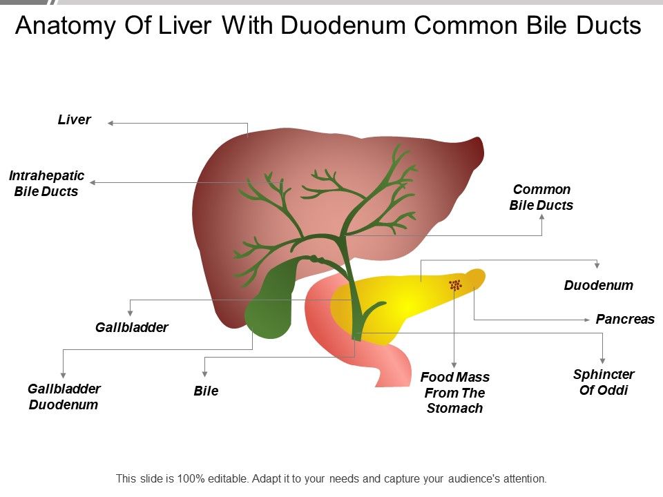
Digestion And Absorption Notes Biology Diagrams From there, the bile ducts in the right lobe flow into the right hepatic duct; bile ducts in the left lobe flow into the left hepatic duct. These, in turn, feed into the common hepatic duct. A branch of the common hepatic duct, the cystic duct, leads to the gallbladder. The gallbladder is a hollow bag which stores and concentrates bile. The liver has two main lobes--the right lobe, which is the largest, and the left lobe. The liver, gallbladder and bile ducts are part of the human the gastrointestinal, or digestive tract. The liver contains many small bile ducts that allow bile that is made by the liver to drain into the gallbladder (see anatomy below).
Bile Ducts Intrahepatic bile ducts in portal area between hepatic lobes, draining hepatic lobules. Extrahepatic bile ducts: Common hepatic duct formed by junction of right and left hepatic bile ducts. Cystic duct connects common hepatic duct with gallbladder. Common bile duct formed by junction of cystic duct and common hepatic duct. Liver Structure: The liver has four lobes which are made up of smaller lobules. The liver's lobules are its functional units. Each lobule is a hexagon shape made up of plates of hepatocytes (special epithelial cells). The plates of hepatocytes radiate outward from a central vein. Through them is a canal where the bile they produce flows outward into the triad's bile duct. The canals are The bile ducts are a series of tubes that drain bile from the liver and either direct it to the gallbladder for temporary storage or pass it into the duodenum where it can be expelled with the feces. The biliary tree as it is known has many different parts, all of which serve the same function, and are prone to a number of diseases that can ultimately affect the liver, gallbladder and/or

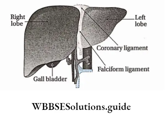
Normal liver anatomy Biology Diagrams
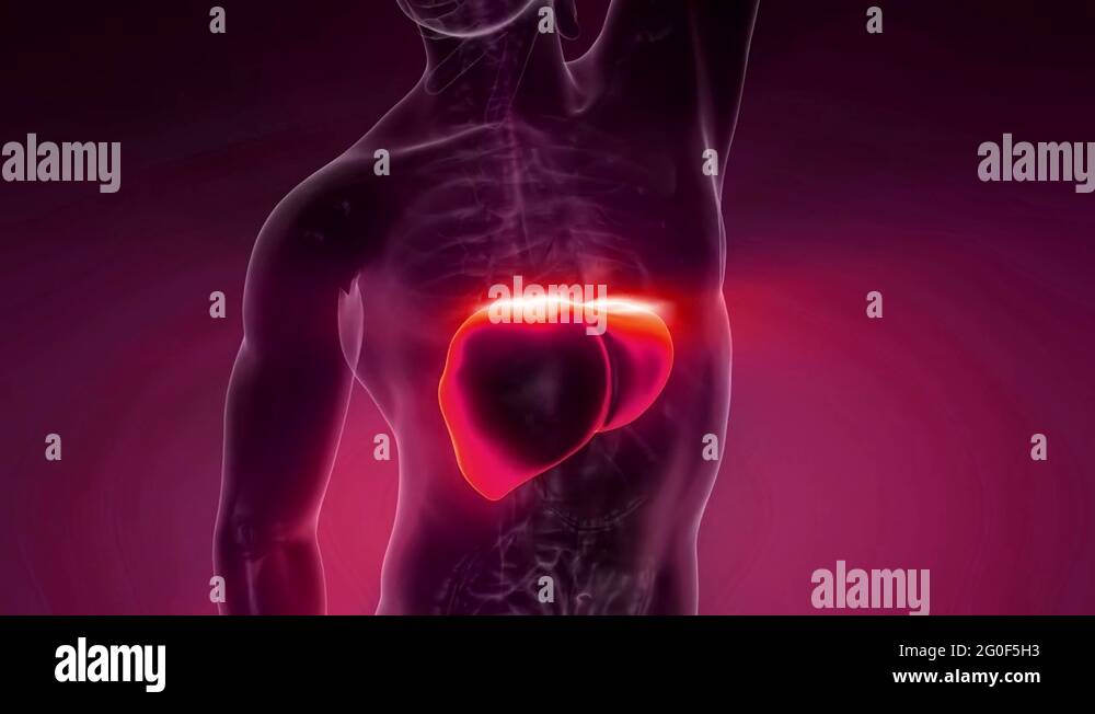
Morphological anatomy of the liver. Based on Couinaud classification, the liver is divided into eight independent functional segments (Figs. 6 and 7). Each segment has its own portal pedicle consisting of the hepatic arterial branch, portal branch, and the bile duct with a separate hepatic venous branch that provides outflow (Fig. 8).
Traditionally, the liver was divided into four anatomical lobes: the right, left, caudate and quadrate lobes. However, this has been superseded by the use of the Couinaud classification which divides the liver into eight functional units (known as segments), supplied by individual segmental hepatic arteries, portal veins and bile ducts, which These bile ducts next join to form the larger left and right hepatic ducts, which carry bile from the left and right lobes of the liver. Those two hepatic ducts join to form the common hepatic duct that drains all bile away from the liver.
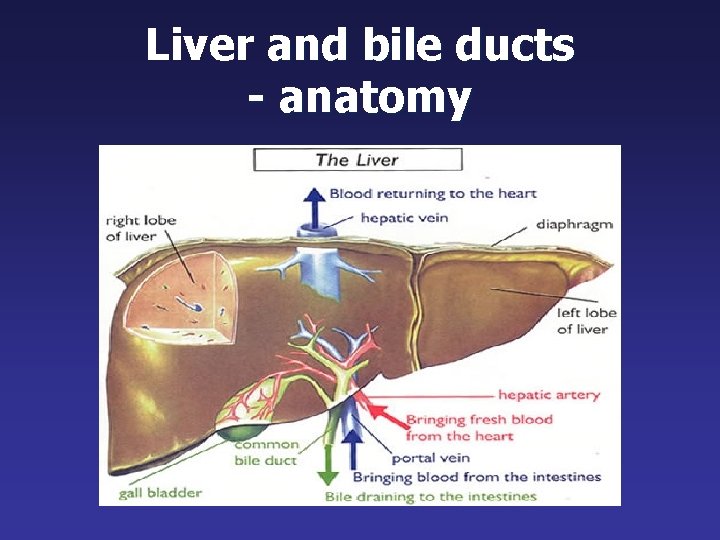
Anatomy of the Liver & Gallbladder Biology Diagrams
As part of the digestive system, the liver creates bile, a fluid that helps digest fats. This bile is stored in the gallbladder, a small pouch beneath the liver, and released into the small intestine when needed for digestion. Liver anatomy consists of two main sections, called lobes, but it's much more than that. This artery then bifurcates into the gastroduodenal artery and the hepatic artery proper, which then also bifurcates into the right and left hepatic arteries. The right and left hepatic arteries mainly supply the non-parenchymal part of the liver, such as the bile ducts within the liver. Now, let's talk about venous drainage of the liver.