
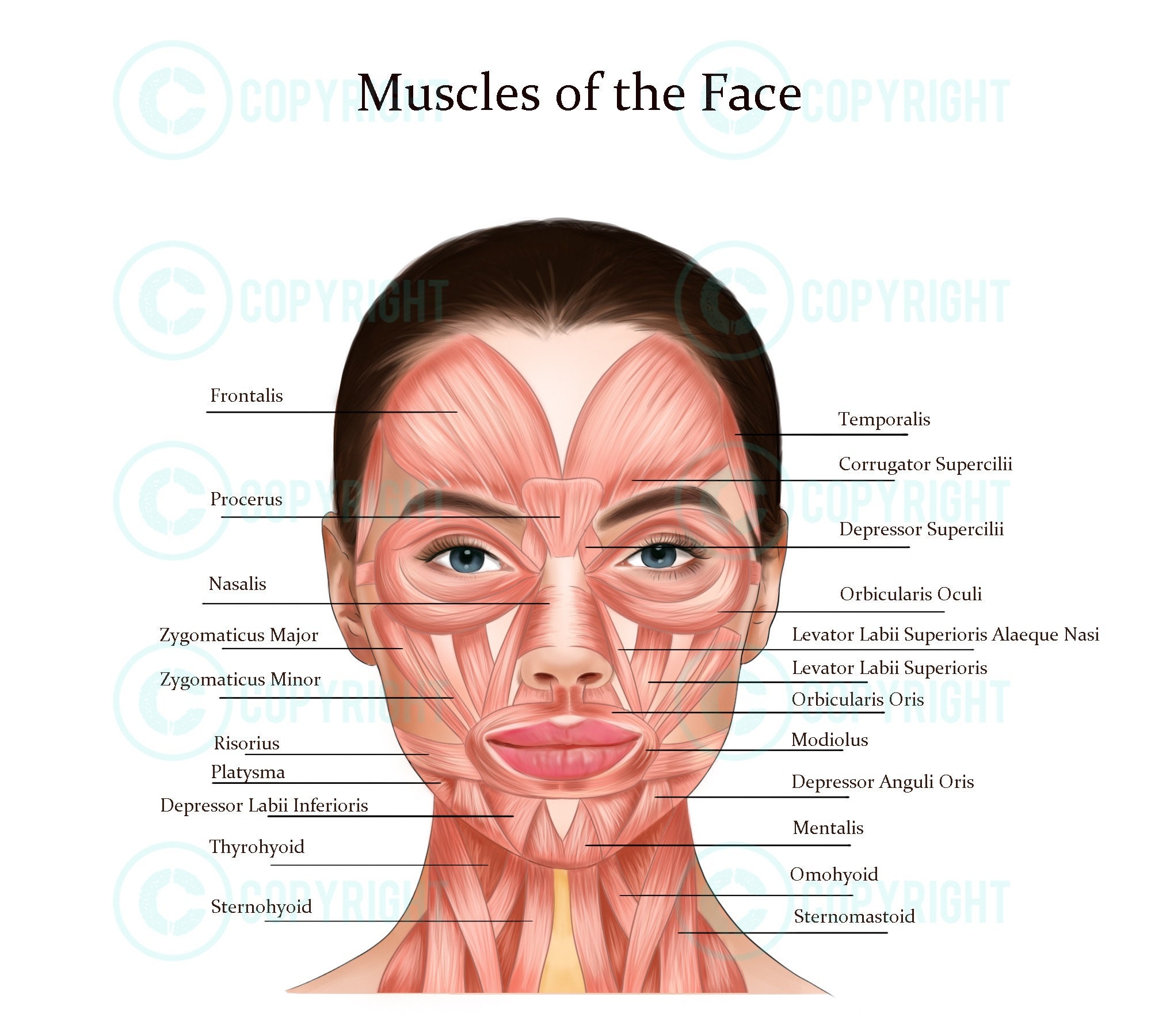
Facial Muscles Diagram Biology Diagrams The following interactive diagram of muscles of human facial expression is an anterior view of some of the muscles and tissues of the head (useful for Indian head massage courses). Click on the pink questions for immediate answers. The muscles / tissues listed below are included in the diagram above. (The numbers are how the muscles are The human face possesses around 30 muscles on each side, depending on how they are counted. The facial muscles are striated muscles that link the facial skin to the skull bone to perform important daily life functions, such as mastication and emotion expression. The facial muscles produce various movements but are often categorized into facial expression (mimetic) and mastication muscles. The Kenhub offers a comprehensive guide to learn the facial muscles anatomy, function and innervation. You can practice with labeled diagrams, interactive quizzes and videos.

Learn about the 20 facial muscles that control your face movements, such as chewing, smiling and talking. Find out what conditions and disorders can affect the facial muscles and how to seek medical attention.
Human face: anatomy, structure and function Biology Diagrams
Learn about the facial muscles, a group of about 20 flat skeletal muscles that produce facial expressions. See diagrams, names, origins, insertions, innervation and blood supply of each muscle group. There are about 20 flat skeletal muscles that construct the facial structure. All of these muscles have different functions in the face. Innervated by the cranial nerve, which is the facial nerve, the muscles control all of our facial expressions. These muscles of the face can be grouped in different categories, depending on their position. Let us have a look at the 20 different muscles of our Nasal Group. The nasal group of facial muscles are associated with movements of the nose and the skin surrounding it.. Nasalis. The nasalis is the largest of the nasal muscles and is comprised of two parts: transverse and alar.. Attachments: Transverse part - originates from the maxilla, immediately lateral to the nose. It attaches onto an aponeurosis across the dorsum of the nose.

2) Occipitalis - not shown on diagram (in back): a. Actions: pulls scalp back b. Innervation: Facial Nerve (Vll) c. Origin: from posterior back of skull d. Insertion: to anteriorly with the galea aponeurotica 3) Orbicularis oculi (sphincter muscle of the eyelid): a. Actions: squinting, closes eye, crowsfeet, eye bags b. Innervation: Facial The facial muscles are also known as the muscles of the facial expression or the mimetic muscles. These muscles are a group of approximately 20 superficial skeletal muscles of the face and scalp divided into five different groups according to their location and function. These groups include: Buccolabial (oral) group: Levator labii superioris, levator labii superioris alaeque nasi, risorius The facial muscles (also called the muscles of facial expression) are situated within the subcutaneous tissue of the face. They are responsible for the movements of skin folds, providing different facial expressions. The facial muscles originate from the bones of the facial skeleton (viscerocranium) and insert into the skin.. These muscles are mostly grouped around the natural orifices of the
