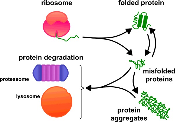
Figure 1 from Degradation of an Old Human Protein Biology Diagrams The half-lives of proteins within cells vary widely, from minutes to several days, and differential rates of protein degradation are an important aspect of cell regulation. Many rapidly degraded proteins function as regulatory molecules, such as transcription factors. The entry of all eukaryotic cells into mitosis is controlled in part by Imbalances in protein levels will occur particularly in protein complexes where the genes encoding individual components of the complex reside on different chromosomes. These imbalances will disrupt protein homeostasis, increase the workload for chaperones, and overload the protein degradation machinery, resulting in a form of toxicity referred Key events in mitosis such as sister chromatid separation and subsequent inactivation of cyclin-dependent kinase 1 are regulated by ubiquitin-dependent proteolysis. These events are mediated by the anaphase-promoting complex (APC), a cell cycle-regulated ubiquitin ligase that assembles multiubiquitin chains on regulatory proteins such as securin and cyclins and thereby targets them for

Mouery et al. utilize deep, quantitative proteomics to reveal a PLK1-regulated program of G2/M protein degradation. They show that the PKA-anchoring protein AKAP2 is degraded in a manner dependent on PLK1 and the E3 ubiquitin ligase SCFβTrCP and that disruption of AKAP2 degradation results in defects of the mitotic cytoskeleton.

Lysosomal degradation ensures accurate chromosomal segregation to ... Biology Diagrams
Xkid is a microtubule plus end-directed motor that moves chromosomes away from spindle poles in early mitosis. Xkid degradation, therefore, facilitates the segregation of chromatids toward spindle poles in anaphase. It is conceivable that Emi1/Rca1 has a role in Cdh1 degradation because Emi1 is an F box protein that interacts with SCF

During mitosis, known degradative functions are mainly restricted to UPS, specialized in ubiquitin-triggered protein degradation and presumably faster than lysosome-dependent degradation. Lysosomes are acidic cytosolic vesicles responsible to enzymatically degrade all types of biological material.

Asymmetric mitosis: Unequal segregation of proteins destined for ... Biology Diagrams
ABSTRACT. Protein expression levels depend on the balance between their synthesis and degradation rates. Even quiescent (G 0) cells display a continuous turnover of proteins, despite protein levels remaining largely constant over time.In cycling cells, global protein levels need to be precisely doubled at each cell division in order to maintain cellular homeostasis, but we still lack a It has recently become clear that several other proteins are degraded at specific points in mitosis. This review (which is part of the Chromosome Segregation and Aneuploidy series) focuses on how specific proteins are selected for proteolysis at defined points in mitosis and how this contributes to the proper coordination of chromosome Since the description of mitosis by Flemming in 1882, studies on somatic cell division have focused on the equal partition of cellular materials, in particular that of the chromosomes and mitotic apparatus, between cell daughters (1, 2).We now report that many mitotic divisions are unequal with respect to pericentrosomal proteins targeted for proteasomal degradation.

