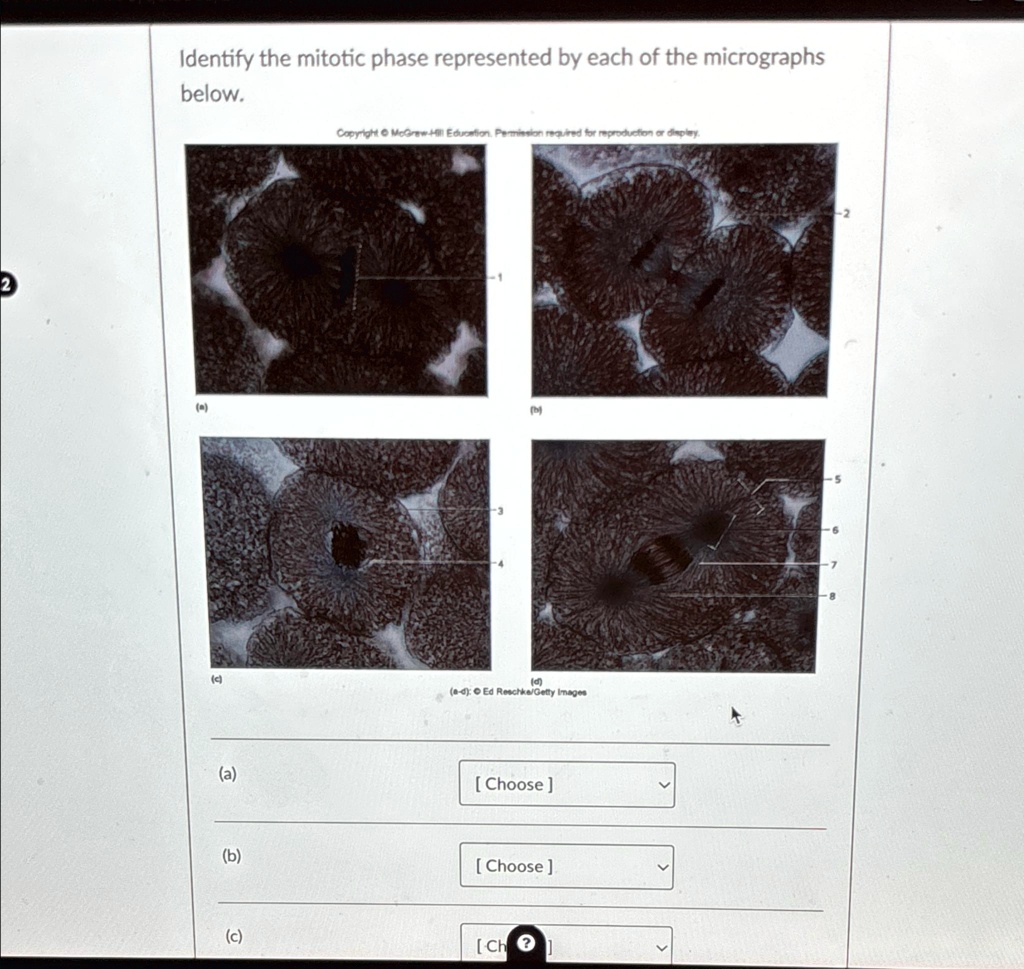
Identify the mitotic phase represented by each of the micrographs below Biology Diagrams Stages of Mitosis Diagram with Labels. The cell synthesizes proteins and organelles needed for the upcoming mitotic phase. It also checks for any errors in DNA replication and repairs any mistakes. Overall, interphase is a critical period for the cell as it allows for growth, DNA replication, and preparation for division.
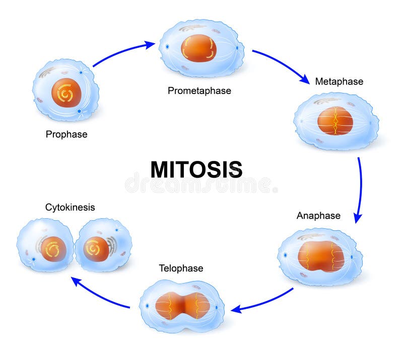
Mitosis Diagram showing the different stages of mitosis. Mitosis is the phase of the cell cycle where the nucleus of a cell is divided into two nuclei with an equal amount of genetic material in both the daughter nuclei. It succeeds the G2 phase and is succeeded by cytoplasmic division after the separation of the nucleus. Explore the stages of mitosis with detailed diagrams. Understand each phase and discover real-world applications of this essential cell division process. Interphase is a part of the cell cycle where the cell copies its DNA as preparation for the M phase (mitotic phase). In interphase, metabolism of the cell increases, and it is often termed

Mitosis: Phases, Applications & Diagrams Explained Biology Diagrams
Before a dividing cell enters mitosis, it undergoes a period of growth called interphase. About 90% of a cell's time in the normal cell cycle may be spent in interphase. G1 phase: The period before the synthesis of DNA. In this phase, the cell increases in mass in preparation for cell division. The G1 phase is the first gap phase. The centrioles complete their duplication during this phase. G2-phase: Second gap or resting phase during which the synthesis of RNA and proteins from the G1 phase continues. During this period, cells store energy as ATP to be utilized during mitosis. At the end of this stage, cells enter the stage of mitotic division. Mitotic Division (M Phase) 1.
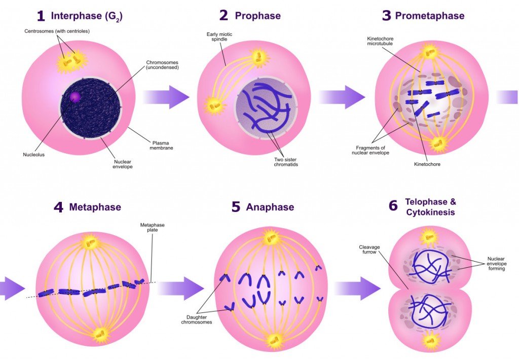
Mitosis succeeds the G2 phase and is followed by cytokinesis where the cytoplasm divides after the separation of the nucleus. It has five phases: prophase, prometaphase, metaphase, anaphase, and telophase. Mitosis forms the basis of sexual reproduction and is important for the growth and development of the embryo. Diagram of Mitosis

Understanding the Process of Mitosis: A Labeled Diagram Biology Diagrams
The stage, or phase, after the completion of mitosis is called interphase. Witness a living plant cell's chromosomes carrying genetic material duplicate during the process of mitosis Time-lapse photography of a live plant cell nucleus undergoing mitosis. (more) See all videos for this article. In this phase, the cell has elongated and is nearly finished dividing. Cell-like features begin to reappear such as the reformation of two nuclei (one for each cell). The chromosomes then de-condense and the mitotic spindle fibres are broken down. Fig 2 - Summary diagram showing the stages of mitosis. Clinical Relevance - Errors of Mitosis. Mitosis Stages Diagram. Mitosis is the process of cell division in which a cell duplicates its genetic material and divides into two daughter cells. This process is essential for the growth, development, and maintenance of multicellular organisms. G1 phase, S phase, and G2 phase. Prophase: During prophase, the chromatin condenses into