Image The lateral sulcus Sylvian fissure separates the temporal lobe Biology Diagrams The temporal lobe, located near the temples, is the second largest lobe of the human cerebrum, accounting for almost one-fourth of the brain's volume. It processes auditory information, forms memories, comprehends language, and regulates emotions through key structures like the hippocampus and primary auditory cortex.

Gross anatomy. The temporal lobe is the second largest lobe, after the larger frontal lobe, accounting 22% of the total neocortical volume 6.. The lobe extends superiorly to the Sylvian fissure, and posteriorly to an imaginary line; the lateral parietotemporal line, which separates the temporal lobe from the inferior parietal lobule of the parietal lobe superiorly and the occipital lobe The temporal lobe is one of the four major lobes of the cerebral cortex. It is the lower lobe of the cortex and has associations with several conditions. Learn more. The temporal lobe is one of the four major lobes of the cerebral cortex in the brain of mammals.The temporal lobe is located beneath the lateral fissure on both cerebral hemispheres of the mammalian brain. [3]The temporal lobe is involved in processing sensory input into derived meanings for the appropriate retention of visual memory, language comprehension, and emotion association.
Temporal Lobe: Definition, Functions, Location & Damage Biology Diagrams
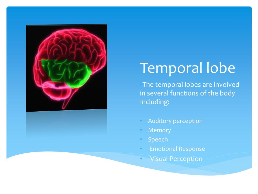
The temporal lobe is visible on the lateral, medial and basal surfaces of the cerebral hemisphere. On the lateral surface, it is demarcated superiorly by the lateral sulcus (or Sylvian fissure) and extends ventrally to the inferior surface of the cerebrum.Posteriorly, the temporal lobe extends to an arbitrary line running between the top of the parietooccipital sulcus and the preoccipital notch. The left temporal lobe is coloured green on this diagram. The temporal lobes are split into three areas: Superior Temporal Gyrus; Middle Temporal Gyrus; Inferior Temporal Gyrus; Near the point where the occipital lobes meet the middle temporal gyrus, is an area called MT, which stands for middle temporal, and is also sometimes called V5.

Learn about the temporal lobe, a part of your brain that helps you use your senses, store and retrieve memories, understand language and process emotions. Find out where it is, what it does and what can go wrong with it. The temporal lobe is in charge of lots of things, including hearing, language, and memory! It makes sense that the temporal lobe helps the brain process sound because it is so close to the ears. Since the temporal lobe is in charge of our sense of hearing, it also plays an important role in music! It helps us process parts of music like rhythm

Temporal Lobe: What It Is, Function, Location & Damage Biology Diagrams
Temporal lobe is located on the sides of the head, above the ears. This region is critical for memory, understanding language, and auditory processing. The hippocampus, part of this lobe, plays a major role in forming new memories. How to Identify and Label the Major Brain Structures on a Diagram. First, locate the cerebrum, the largest

Temporal Lobe: The brains contain four lobes in the cortex, including the occipital, parietal, temporal, and frontal lobes. The temporal lobe located just beneath the lateral fissure and crisscrossing both fissures of the brain. This vital structure of the temporal lobe supports process the sensory input, including pain and the auditory stimuli. It further helps you […]