Learn anatomy Biology Diagrams The spinal cord is a tubular structure composed of nervous tissue that extends from the brainstem and continues distally before tapering at the lower thoracic/upper lumbar region as the conus medullaris. The spinal cord is anchored distally by the filum terminale, a fibrous extension of the pia mater anchoring the spinal cord to the coccyx.[1] Protecting the spinal cord is the surrounding Learn about the spinal cord, a tubular bundle of nervous tissue that connects the brain and the peripheral nervous system. Explore its structure, membranous coverings, blood supply, and clinical relevance.
Learn about the structure, function and blood supply of the spinal cord, a column of nerve tissue that connects the brain and the periphery. Find out how the spinal cord is divided into regions, tracts, nerves and reflexes.
Spinal cord: Anatomy, functions, and injuries Biology Diagrams
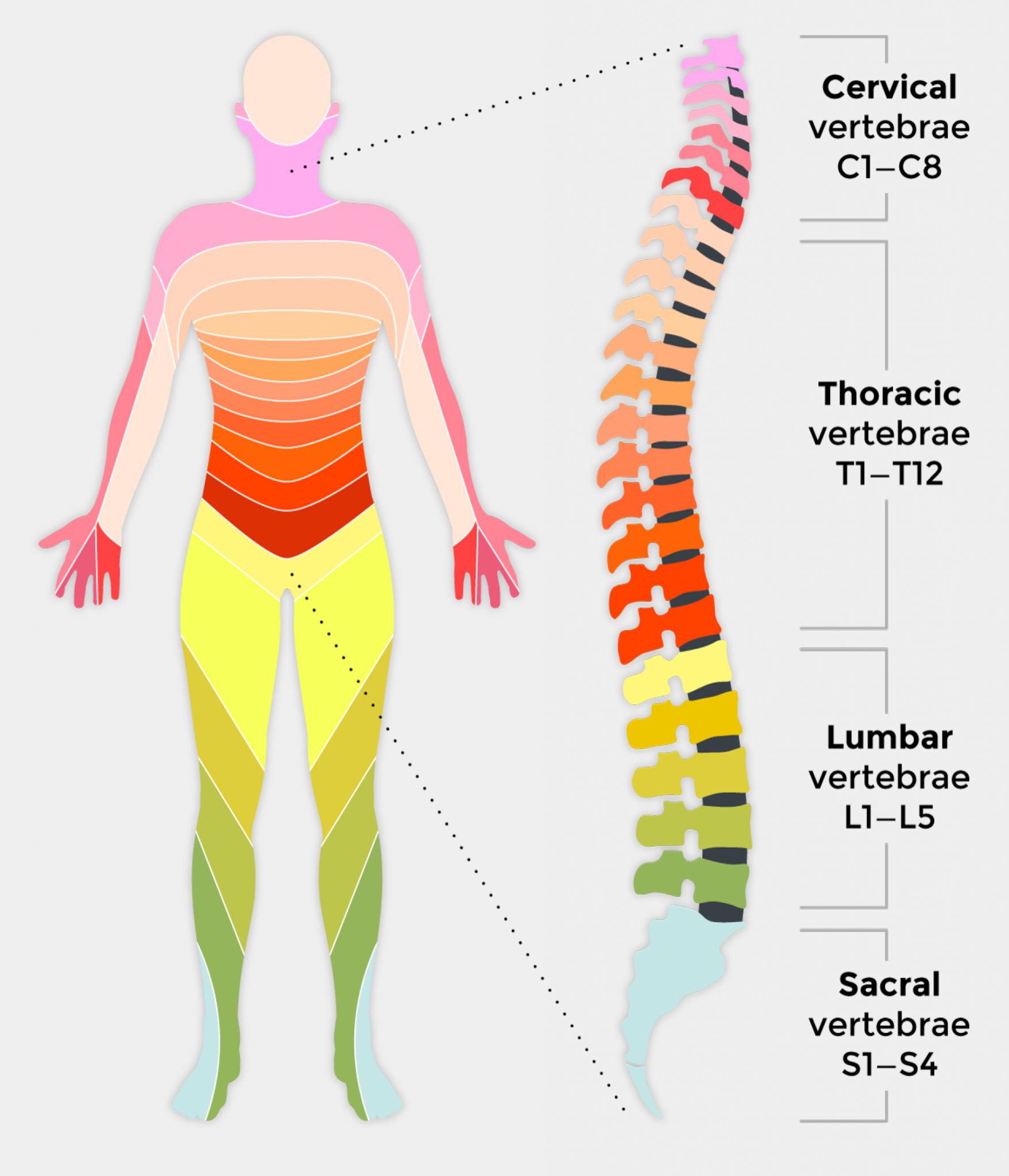
Learn about the spinal cord, a part of the central nervous system that conveys information between the brain and the body. Explore its segments, nerves, meninges, blood supply, and tracts with diagrams, videos, quizzes and more. The spinal cord study is one of the most complex yet quite a fascinating part of the nervous system. Its complex connections, the development defects, the lesions, and clinical presentation are quite overwhelming and warrants a better understanding of its anatomical and physiological nature. This topic has received extensive study and revealed many minute details. Learn about the spinal cord, a long, cylindrical structure that transmits information between the brain and the body. Find out its location, regions, meninges, internal structure, spinal nerves, and clinical relevance.
Learn about the spinal cord, a tube of tissue that carries nerve signals from your brain to the rest of your body and back. Find out how it works, where it's located, what nerves are in it and what conditions can affect it.
Spinal Cord Anatomy Biology Diagrams
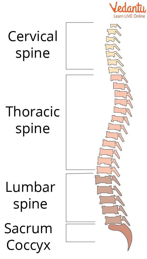
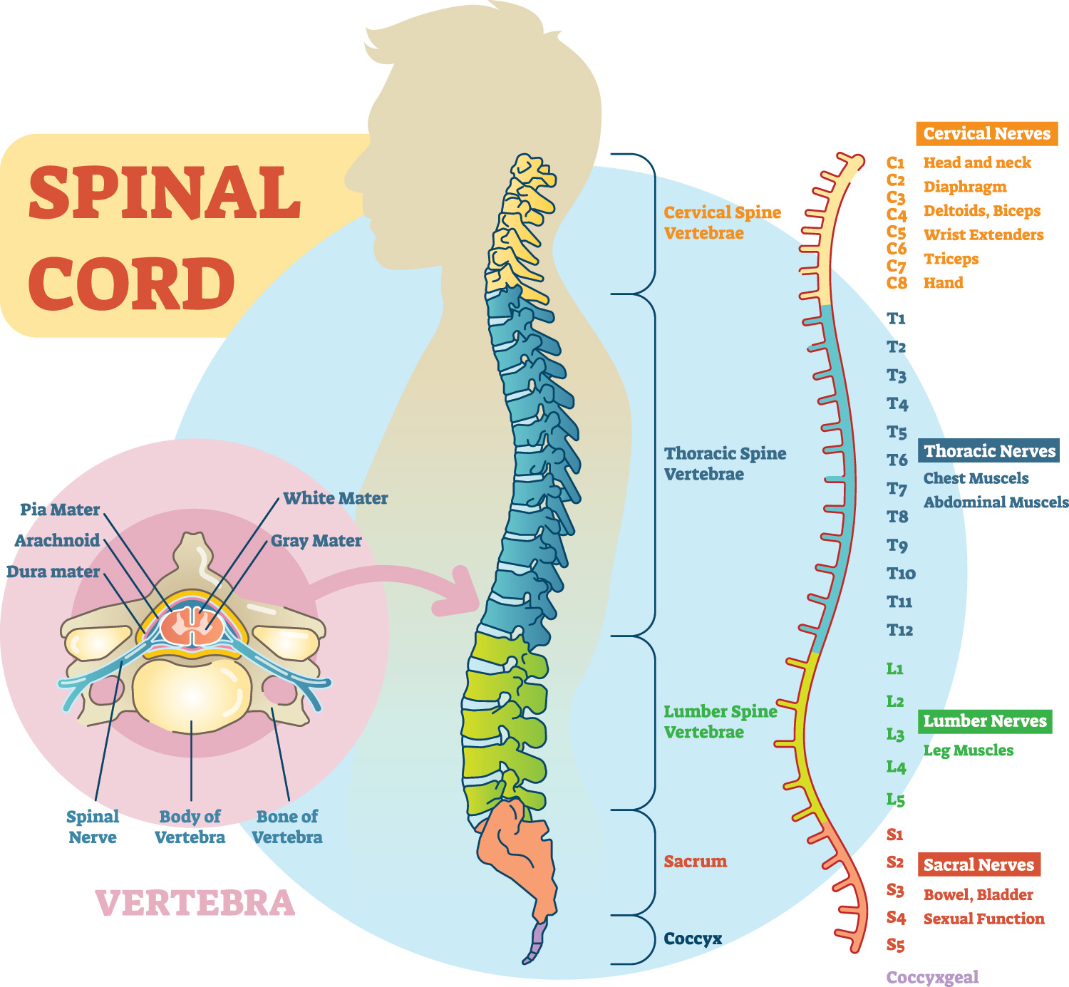
The spinal cord is part of the central nervous system (CNS), which extends caudally and is protected by the bony structures of the vertebral column. It is covered by the three membranes of the CNS, i.e., the dura mater, arachnoid and the innermost pia mater. In most adult mammals it occupies only the upper two-thirds of the vertebral canal as the growth of the bones composing the vertebral
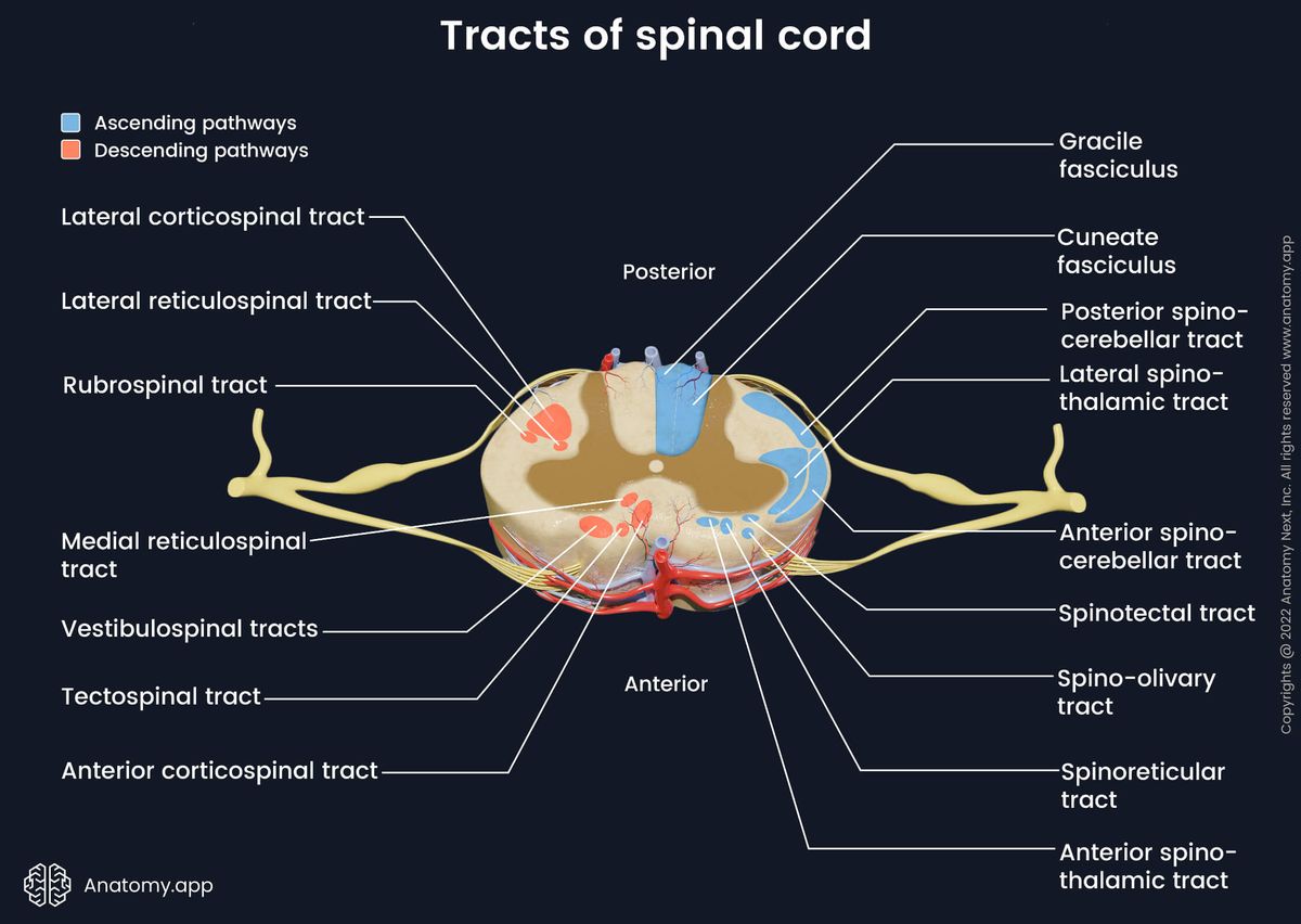
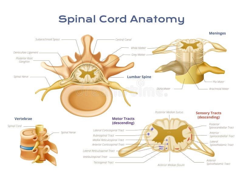
Learn about the spinal cord's structure, location, and role in carrying signals between the brain and the body. Find out how spinal cord injuries can affect movement and sensation and what treatments are available. Learn about the regions, horns, columns, and tracts of the spinal cord, the major organ of the central nervous system. See how the spinal cord connects with the brain, the periphery, and the autonomic nervous system.