
Lumbar Plexus Anatomy Biology Diagrams Lumbosacral Plexus Diagram. Move the Slider to Practice! The lumbosacral plexus is a network of nerve fibers that innervates muscles and provides sensation from the lower limbs. It is formed by the anterior rami of T12-L4/5 nerve roots. To simplify, the lumbar (T12-L4/5) and sacral plexuses (L4-S4) will be discussed separately. The lumbar plexus is a complex network of nerves formed by the anterior rami of the lumbar spinal nerves L1 through L4, sometimes with contributions from T12. This plexus gives rise to several important nerves that innervate the lower abdomen, pelvis, and lower extremities.[5] Structure Components: The lumbar plexus consists of the anterior (ventral) rami The lumbar plexus is a complex neural network formed by the lower thoracic and lumbar ventral nerve roots (T12 to L5) which supplies motor and sensory innervation to the lower limb and pelvic girdle. Summary origin: ventral rami of T12 to L5 c
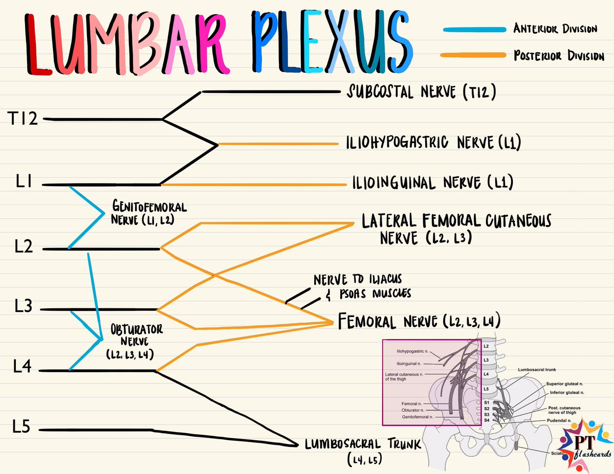
The lumbar plexus is a network of nerve fibres that supplies the skin and musculature of the lower limb.. It is located in the lumbar region, within the substance of the psoas major muscle and anterior to the transverse processes of the lumbar vertebrae.. The plexus is formed by the anterior rami (divisions) of the lumbar spinal nerves L1, L2, L3 and L4. . It also receives contributions from

Anatomy, Diagram, Location, Functions Biology Diagrams
The lumbar plexus is a web of nerves (a nerve plexus) in the lumbar region of the body which forms part of the larger lumbosacral plexus.It is formed by the divisions of the first four lumbar nerves (L1-L4) and from contributions of the subcostal nerve (T12), which is the last thoracic nerve.Additionally, the ventral rami of the fourth lumbar nerve pass communicating branches, the lumbosacral The lumbosacral plexus refers to the combination of the lumbar plexus and the sacral plexus. It is derived from the L2, L3, L4, L5, S1, and S2 roots and is involved in nerve conduction in the lower back and pelvic region. Simplified coronal diagram of lumbosacral plexus, depicted on a background of psoas, iliacus and piriformis muscles The Lumbar Plexus (*136 (plexus lumbalis) (Figs. 822, 823, 824). 826- Diagram of segmental distribution of the cutaneous nerves of the right lower extremity. Front view. The Genitofemoral Nerve (n. genitofemoralis; genitocrural nerve) arises from the first and second lumbar nerves. It passes obliquely through the substance of the Psoas
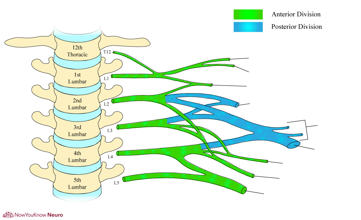
The lumbar plexus is a network of nerves that arises from the anterior rami of spinal nerves L1-L4, along with a contribution from the anterior ramus of spinal nerve T12.It is located on the posterior abdominal wall, anterior to the transverse processes of the lumbar vertebrae and within the posterior portion of the psoas major muscle.. The lumbar plexus gives rise to several branches which

Lumbar plexus: Anatomy, branches and innervation Biology Diagrams
The lumbosacral plexus is a network of nerve fibers, derived from the roots of lumbar and sacral spinal nerves that branch out to form the nerves supplying the lower limb.In the human body, there are 31 pairs of spinal nerves corresponding to a segment of the vertebral column: cervical (C1-C8), thoracic (T1-T12), lumbar (L1-L5), sacral (S1-S5), and coccygeal.

The anterior divisions of the lumbar nerves, sacral nerves, and coccygeal nerve form the lumbosacral plexus, the first lumbar nerve being frequently joined by a branch from the twelfth thoracic. For descriptive purposes this plexus is usually divided into three parts:
