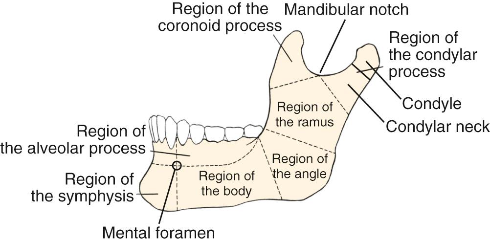
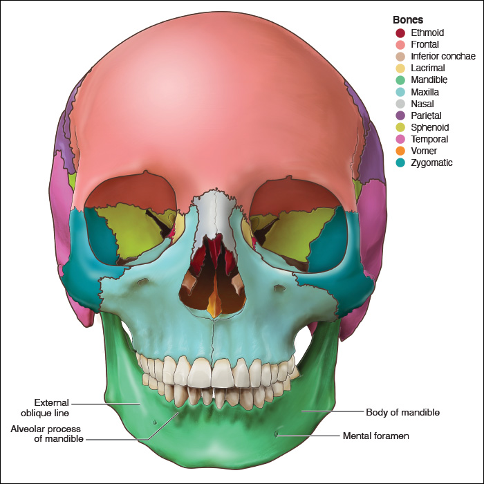
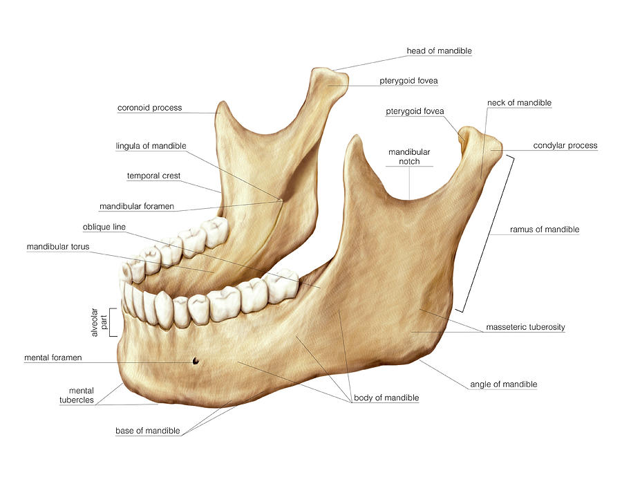
Mandibula E Maxila Anatomia Biology Diagrams The temporomandibular joint (TMJ) is formed by the articulation of the mandible and the temporal bone of the cranium. It is located anteriorly to the tragus of the ear, on the lateral aspect of the face. In this article, we shall look at the anatomy of the temporomandibular joint - its articulating surfaces, ligaments and clinical correlations. The mandible is the largest bone in the human skull, forming the lower jawline and shaping the contour of the inferior third of the face (see Image. Mandible Anatomy).[1] Articulation with the skull base at the bilateral temporomandibular joints allows a range of movements facilitated by associated muscles, including dental occlusion with the maxilla (see Image. Jaw Anatomy, Lateral View). The
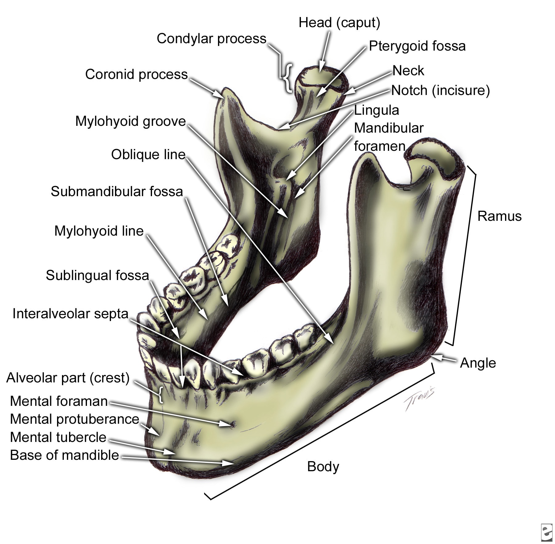
A synovial joint that enables the intricate movements required for life is the temporomandibular joint (TMJ), also known as the jaw joint. This joint is between the temporal bone's mandibular fossa and the mandibular condylar head. This system, which includes the TMJ, teeth, and soft tissue, is involved in speech, eating, and breathing. The temporomandibular joint (TMJ), or jaw joint, is a synovial joint that allows the complex movements necessary for life. It is the joint between condylar head of the mandible and the mandibular fossa of the temporal bone. GA, Retrodiscal Tissue of the Temporomandibular Joint: Clinical Anatomy and its Role in Diagnosis and Treatment of The TMJ is a unique joint involved in a number of important functions, including mastication and speech, 1 but more simply, it allows the articulation between the upper and lower jaws. 2 This
Radiology Reference Article - Radiopaedia.org Biology Diagrams
The temporomandibular joint (TMJ) is involved in mastication (chewing) and speech and it is one of the most frequently moved joints in humans.[1] This synovial joint must be able to respond to significant biomechanical load.[2] It is made up of the articulating surface of the temporal bone and the head of the mandible. The whole joint is enclosed in a fibrous capsule.[3] The temporomandibular joint (TMJ) is an atypical synovial joint located between the condylar process of the mandible and the mandibular fossa and articular eminence of the temporal bone. It is divided into a superior discotemporal space and inferior discomandibular space by the TMJ disc (or meniscus).
In anatomy, the temporomandibular joints (TMJ) are the two joints connecting the jawbone to the skull. The lower joint compartment formed by the mandible and the articular disc is involved in rotational movement—this is the initial movement of the jaw when the mouth opens. The upper joint compartment formed by the articular disc and the The temporomandibular joint (TMJ) is a diarthrosis, better defined as a ginglymoarthrodial joint. TMJ is composed of a synovial cavity, articular cartilage and a capsule that covers the same joint. We find the synovial fluid and several ligaments. The joint is the union of the temporal bone cavity with the mandibular condyle. Anatomy