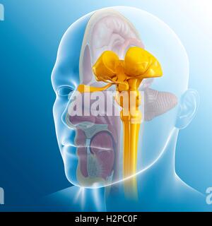
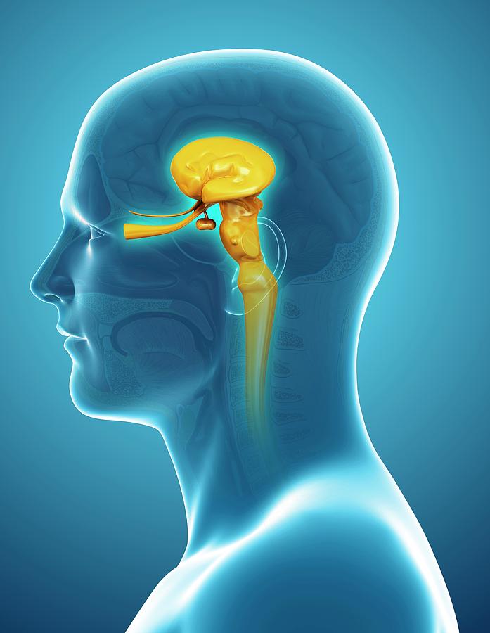
Neuroanatomy Mesencephalon Midbrain Article Biology Diagrams The brainstem, including the midbrain, the pons, and the medulla, comprises several nerves, pathways, reflex centers, and nuclei (see Image. Midbrain Anatomy). The midbrain is the smallest portion of the brainstem (about 1.5 cm) and its most cranial structure. It is in the brainstem between the pons caudally (mesencephalic-pons groove) and the diencephalon, which includes the thalamus, the midbrain, region of the developing vertebrate brain that is composed of the tectum and tegmentum. The midbrain serves important functions in motor movement, particularly movements of the eye, and in auditory and visual processing.It is located within the brainstem and between the two other developmental regions of the brain, the forebrain and the hindbrain; compared with those regions, the

The midbrain (also known as the mesencephalon) is the most superior of the three regions of the brainstem. It acts as a conduit between the forebrain above and the pons and cerebellum below. In this article, we will discuss the anatomy of the midbrain - its external anatomy, internal anatomy, and vasculature. 712- Transverse section of mid-brain at level of superior colliculi. Structure of the Cerebral Peduncles (Figs. 711, 712). —On transverse section, each peduncle is seen to consist of a dorsal and a ventral part, separated by a deeply pigmented lamina of gray substance, termed the substantia nigra. The midbrain, also known as the mesencephalon, is the smallest, most cranial part of the brainstem. It is located between the diencephalon (which includes the thalamus and hypothalamus) and the pons.On a transverse section, midbrain can be divided into three main regions: the crus cerebrum, the tegmentum (the ventral part) and the tectum (the dorsal part).To demarcate the boundaries between
The Midbrain: Anatomy, Function, and Treatment Biology Diagrams
The midbrain or mesencephalon is the uppermost portion of the brainstem connecting the diencephalon and cerebrum with the pons. [2] It consists of the cerebral peduncles, tegmentum, and tectum.. It is functionally associated with vision, hearing, motor control, sleep and wakefulness, arousal (), and temperature regulation.[3]The name mesencephalon comes from the Greek mesos, "middle", and Gross anatomy The midbrain lies between the thalamus (rostrally) and pons (caudally). Lateral sides of the midbrain are covered and hidden by the hippocampal gyri of the brain.. Anterior (ventral) surface. The anterior surface of the midbrain is marked by the two stalks called the cerebral peduncles.The peduncles are composed of many pathways that travel between the cerebral cortex and spinal Tegmentum: This anterior surface of the midbrain contains numerous structures including the reticular formation, the periaqueductal gray (PAG) matter, certain cranial nerve nuclei, sensory and motor nerve pathways (the corticospinal and spinothalamic tract), the red nucleus, the substantia nigra, and the ventral tegmental area (VTA).; Tectum: The posterior surface of the midbrain contains the

The most important function of the midbrain is controlling visual reflexes, like pupil size. When we look at a patient's pupils with a flashlight, we are assessing the function of the midbrain. Nuclei of the Midbrain Tegmentum. The midbrain is divided into a tegmentum (Latin, "covering") and a tectum (Latin, "roof").