scienceallthetime Blood Circulation in Human Body Biology Diagrams Arteries make up a major part of the circulatory system, with the veins and heart being the other main components. Arteries make up tubelike structures responsible for transporting fluid (i.e., blood for the circulatory system and lymph for the lymphatic system) to and from every organ in the body. Mainly, arteries manage the transportation of oxygen, nutrients, and hormones through our bodies
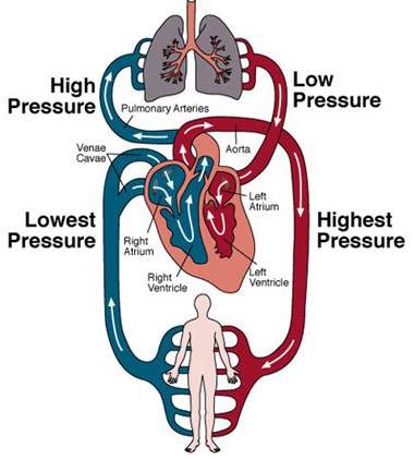
Your circulatory system, or cardiovascular system, supplies oxygen and nutrients to your whole body and removes waste through your blood. Your heart pumps blood that flows through your arteries, veins and capillaries. These blood vessels and your heart form your circulatory system. They work together to ensure your cells have what they need. The aorta, the largest artery in our body, exemplifies this type. - Muscular Arteries: Found farther from the heart, these arteries contain more smooth muscle tissue and fewer elastic fibers. They regulate blood flow to specific organs and regions. ### 2. Artery Wall Layers. The walls of arteries consist of three distinct layers:
List of arteries of the human body Biology Diagrams
These are tiny blood vessels between arteries and veins that distribute oxygen-rich blood to the body. When the heart pumps, blood moves through the circulatory system. Blood leaving the heart through the arteries is full of oxygen. The arteries branch off into smaller and smaller tubes. These bring oxygen and other nutrients to the cells of Arteries, part of your circulatory (cardiovascular) system, are the blood vessels that bring oxygen-rich blood from your heart to all of your body's cells. They play a crucial role in distributing oxygen, nutrients and hormones throughout your body. Arteries keep your body alive and healthy by delivering what your cells and tissues need. As arteries extend further from the heart, they usually become narrower. Additionally, arteries will continually branch off into smaller arteries, arterioles and capillaries, forming the vascular bed. The major arteries in the body are: The aorta. The largest artery in the body, which connects directly to the left ventricle of the heart.

This is a list of arteries of the human body.. The aorta; The arteries of the head and neck. The common carotid artery. The external carotid artery; The triangles of the neck; The internal carotid artery; The arteries of the brain; The arteries of the upper extremity The subclavian artery; The axilla. The axillary artery; The brachial artery; The radial artery; The ulnar artery

Arteries: What They Are, Anatomy & Function Biology Diagrams
The first aortic branches are your coronary arteries, which nourish your heart muscle. Many more arteries branch off your aorta as it travels through your chest and belly. Ultimately, your aorta splits (bifurcates) into two terminal branches called your iliac arteries. Your aorta and your iliac arteries form an upside-down Y near your belly button. The blood circulatory system (cardiovascular system) delivers nutrients and oxygen to all cells in the body. It consists of the heart and the blood vessels running through the entire body. The arteries carry blood away from the heart; the veins carry it back to the heart. The system of blood vessels resembles a tree: The "trunk" - the main artery (aorta) - branches into large arteries The intercostal arteries are a pair of arteries on either side of the body that send blood to various areas of the torso, including the vertebrae, spinal cord, back muscles, and skin. Superior