
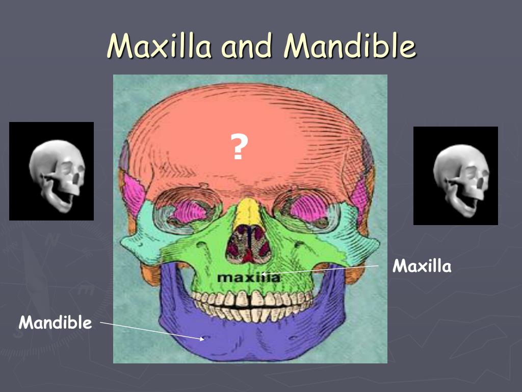
Skeletal System PowerPoint Presentation Biology Diagrams The maxilla is the most important bone of the midface. It has a central location and provides structural support to the viscerocranium. It has functional and aesthetic significance as it has a fundamental role in facial architecture, separates the nasal and oral cavities, forms the upper jaw, and contains the maxillary sinus (See Image. Maxillae).[1][2] In vertebrates, the maxilla (pl.: maxillae / m æ k ˈ s ɪ l iː /) [2] is the upper fixed (not fixed in Neopterygii) bone of the jaw formed from the fusion of two maxillary bones. In humans, the upper jaw includes the hard palate in the front of the mouth. [3] [4] The two maxillary bones are fused at the intermaxillary suture, forming the anterior nasal spine.This is similar to the mandible The maxilla is sometimes called the upper jaw, usually in relation to the dentition. Gross anatomy. The body of the maxilla is roughly pyramidal and has four surfaces that surround the maxillary sinus, the largest paranasal sinus: anterior, infratemporal (posterior), orbital and nasal. It also has four processes: zygomatic, frontal, alveolar
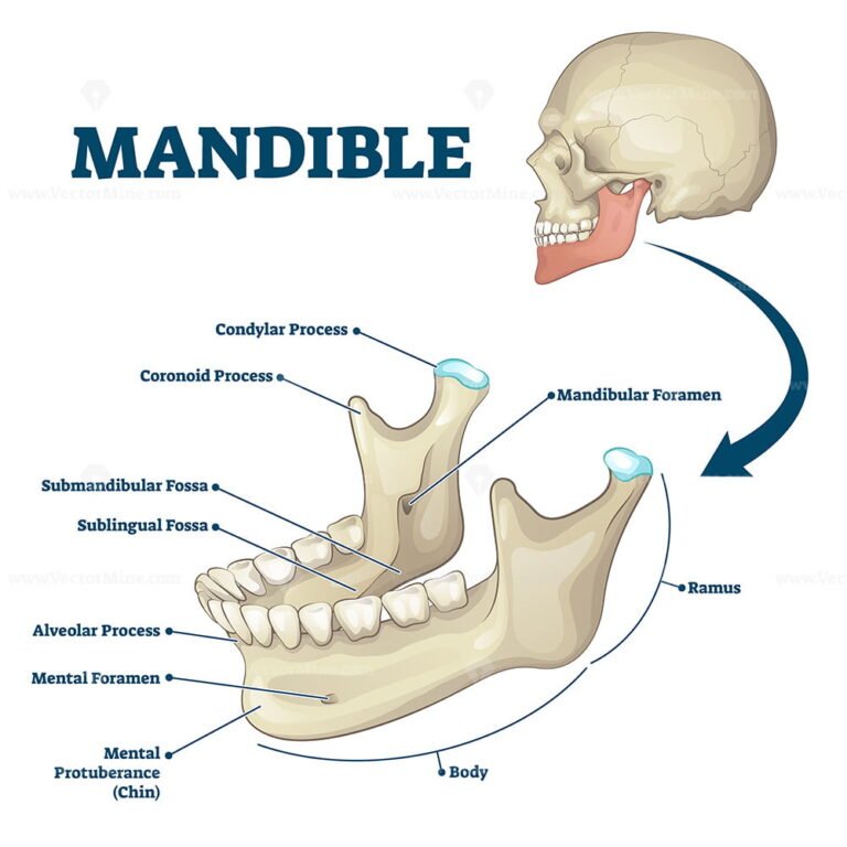
The mandible bone's function is to open and close the mouth for chewing and speech. It also helps protect the organs of the face and hold the lower set of teeth in place. The mandible and the maxilla form the lower and upper parts of the jaw, respectively. The mandible is attached to the major muscle groups of mastication (chewing)
Anatomy, Head and Neck, Maxilla Biology Diagrams
The mandible is the largest bone in the human skull, forming the lower jawline and shaping the contour of the inferior third of the face (see Image. Mandible Anatomy).[1] Articulation with the skull base at the bilateral temporomandibular joints allows a range of movements facilitated by associated muscles, including dental occlusion with the maxilla (see Image. The maxilla is a bone which helps to make up the skull. It is specifically located in the mid face, forms the upper jaw, separates the nasal and oral cavities, and contains the maxillary sinuses (located on each side of the nose. Maxilla (plural: maxillae) is one of the eight facial bones that form the facial skeleton. It is the second largest bone of the face. As it forms the upper jaw holding the upper set of teeth, it is sometimes referred to as the upper jaw bone. It also forms the lower parts of eye sockets and nasal cavities.
The mandible, located inferiorly in the facial skeleton, is the largest and strongest bone of the face.. It forms the lower jaw and acts as a receptacle for the lower teeth. It also articulates on either side with the temporal bone, forming the temporomandibular joint.. In this article, we will look at the anatomy and clinical importance of the mandible. It forms the anterior part of the skull and includes the mandible, maxilla, frontal bone, nasal bones, and zygoma as the primary bones. Facial bone anatomy is complex yet elegant in its suitability to serve a multitude of functions. The image below provides an overview of the anterior features of the skull.

Location, Functions, Anatomy, & Diagram Biology Diagrams
Maxilla. The maxilla, also known as the upper jaw, is a vital viscerocranium structure of the skull.It is involved in the formation of the orbit, nose and palate, holds the upper teeth and plays an important role for mastication and communication.. This bone consists of five major parts, one being the body and four being projections named processes (frontal, zygomatic, palatine, alveolar).
