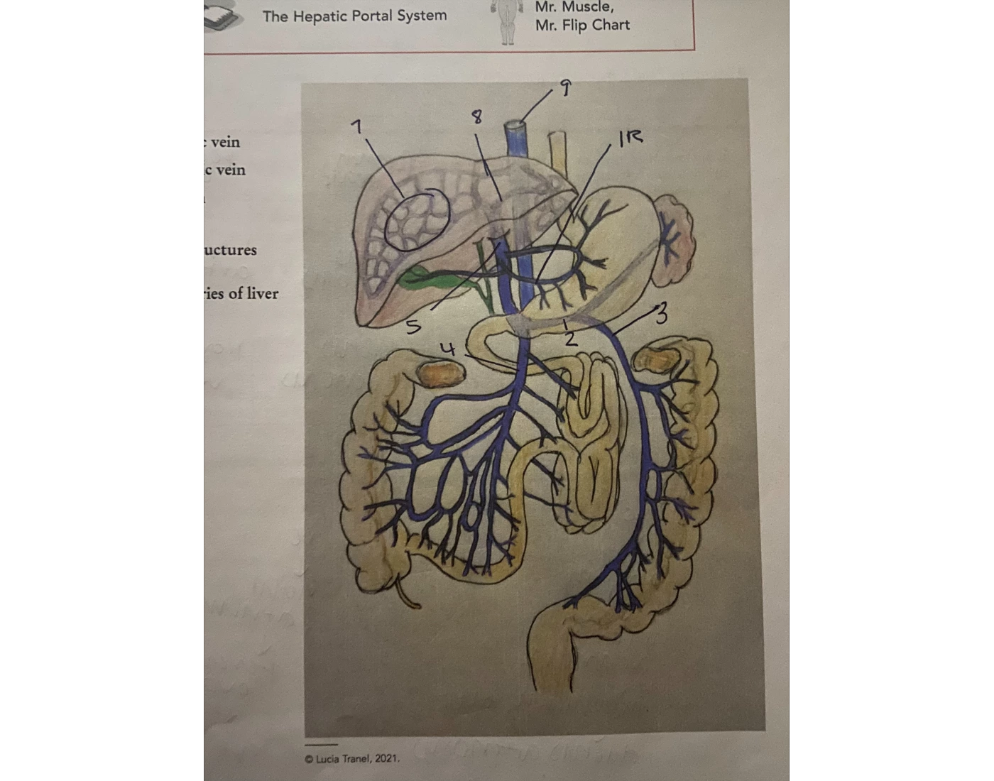
The Cardiovascular System Biology Diagrams Large veins that are considered part of the portal venous system are the: . Hepatic portal vein; Splenic vein; Superior mesenteric vein; Inferior mesenteric vein; The superior mesenteric vein and the splenic vein come together to form the actual hepatic portal vein.The inferior mesenteric vein connects in the majority of people on the splenic vein, but in some people, it is known to connect on The hepatic portal vein is an important and unique vein that receives blood from structures of the abdomen and transports it into the liver for filtration and processing. This vein is part of the hepatic portal system that receives all of the blood draining from the abdominal digestive tract, as well as from the pancreas, gallbladder, and spleen.
The hepatic portal system is the system of veins comprising the hepatic portal vein and its tributaries. The liver consumes about 20% of total body oxygen when at rest, so the total liver blood flow is quite high. Blood flow to the liver is unique in that it receives both oxygenated and partially deoxygenated blood.

Anatomy, Abdomen and Pelvis, Portal Venous System (Hepatic Portal ... Biology Diagrams
The portal vein, or hepatic portal vein, is the main blood vessel of the portal venous system (PVS), which delivers blood to the liver from the stomach, intestines, spleen, gallbladder, and pancreas. Patients with refractory ascites or GI bleeding may benefit from the surgical placement of shunts between the portal and systemic circulation

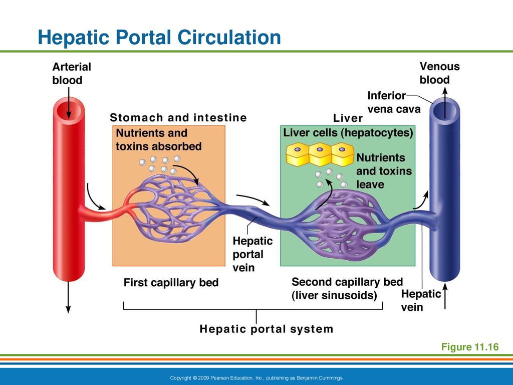
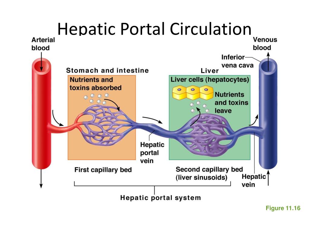
The hepatic portal system is the system of veins that transports blood from the digestive tract to the liver. It consists of the hepatic portal vein and other veins that drain into the hepatic portal vein, viz. the superior mesenteric vein, the inferior mesenteric vein, and the splenic vein. Your portal vein, sometimes called your hepatic portal vein, carries blood into your liver. Meanwhile, your hepatic veins carry blood out of your liver. Your portal vein is part of your portal venous system. This is a network of veins that drains blood from organs in your belly and sends it to your liver for processing. The veins that drain the gastrointestinal organs parallel the major arteries that supply the foregut, midgut, and hindgut, including the celiac, superior mesenteric, and the inferior mesenteric arteries respectively. These veins eventually convene at the portal vein, forming a single venous inflow tract into the liver. The celiac vein drains the foregut structures, including the stomach
Portal Vein Function, Location, and Anatomy Biology Diagrams
The portal vein, formed by the mesenteric and splenic veins, supplies 70% of the blood to the liver. Blood from the portal vein blood contains absorbed nutrients (such as glucose, fatty acids and amino acids) from the GI tract. Firstly, oxygen is delivered to the hepatic circulation from both of these arteries.
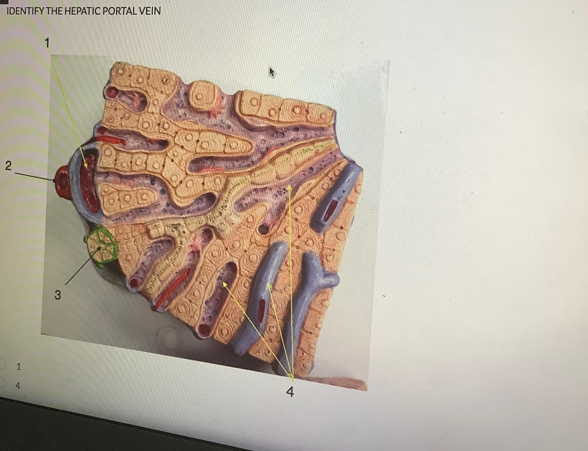
Note enlargement of the hepatic vein due to increased flow directly drained from the PVS to its lumen, skipping the hepatic parenchyma. Other iatrogenic findings may result from post-surgical changes. Liver surgery mainly consists in anatomical-oriented resections, based on the intrahepatic distribution of portal branches and hepatic veins .